The role of bone scanning in severe frostbite of the feet in a
Anuncio
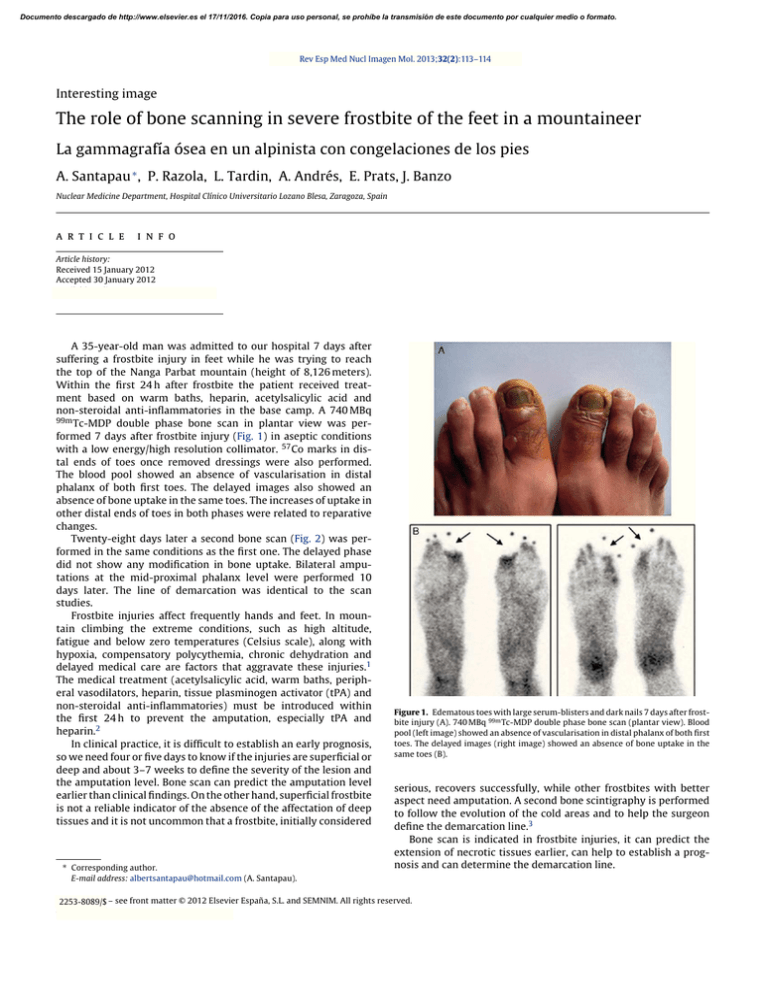
Documento descargado de http://www.elsevier.es el 17/11/2016. Copia para uso personal, se prohíbe la transmisión de este documento por cualquier medio o formato. Rev Esp Med Nucl Imagen Mol. 2013;32(2):113–114 Interesting image The role of bone scanning in severe frostbite of the feet in a mountaineer La gammagrafía ósea en un alpinista con congelaciones de los pies A. Santapau ∗ , P. Razola, L. Tardin, A. Andrés, E. Prats, J. Banzo Nuclear Medicine Department, Hospital Clínico Universitario Lozano Blesa, Zaragoza, Spain a r t i c l e i n f o Article history: Received 15 January 2012 Accepted 30 January 2012 Available online xxx A 35-year-old man was admitted to our hospital 7 days after suffering a frostbite injury in feet while he was trying to reach the top of the Nanga Parbat mountain (height of 8,126 meters). Within the first 24 h after frostbite the patient received treatment based on warm baths, heparin, acetylsalicylic acid and non-steroidal anti-inflammatories in the base camp. A 740 MBq 99m Tc-MDP double phase bone scan in plantar view was performed 7 days after frostbite injury (Fig. 1) in aseptic conditions with a low energy/high resolution collimator. 57 Co marks in distal ends of toes once removed dressings were also performed. The blood pool showed an absence of vascularisation in distal phalanx of both first toes. The delayed images also showed an absence of bone uptake in the same toes. The increases of uptake in other distal ends of toes in both phases were related to reparative changes. Twenty-eight days later a second bone scan (Fig. 2) was performed in the same conditions as the first one. The delayed phase did not show any modification in bone uptake. Bilateral amputations at the mid-proximal phalanx level were performed 10 days later. The line of demarcation was identical to the scan studies. Frostbite injuries affect frequently hands and feet. In mountain climbing the extreme conditions, such as high altitude, fatigue and below zero temperatures (Celsius scale), along with hypoxia, compensatory polycythemia, chronic dehydration and delayed medical care are factors that aggravate these injuries.1 The medical treatment (acetylsalicylic acid, warm baths, peripheral vasodilators, heparin, tissue plasminogen activator (tPA) and non-steroidal anti-inflammatories) must be introduced within the first 24 h to prevent the amputation, especially tPA and heparin.2 In clinical practice, it is difficult to establish an early prognosis, so we need four or five days to know if the injuries are superficial or deep and about 3–7 weeks to define the severity of the lesion and the amputation level. Bone scan can predict the amputation level earlier than clinical findings. On the other hand, superficial frostbite is not a reliable indicator of the absence of the affectation of deep tissues and it is not uncommon that a frostbite, initially considered ∗ Corresponding author. E-mail address: albertsantapau@hotmail.com (A. Santapau). Figure 1. Edematous toes with large serum-blisters and dark nails 7 days after frostbite injury (A). 740 MBq 99m Tc-MDP double phase bone scan (plantar view). Blood pool (left image) showed an absence of vascularisation in distal phalanx of both first toes. The delayed images (right image) showed an absence of bone uptake in the same toes (B). serious, recovers successfully, while other frostbites with better aspect need amputation. A second bone scintigraphy is performed to follow the evolution of the cold areas and to help the surgeon define the demarcation line.3 Bone scan is indicated in frostbite injuries, it can predict the extension of necrotic tissues earlier, can help to establish a prognosis and can determine the demarcation line. 2253-654X/$ 2253-8089/$ – see front matter © 2012 Elsevier España, S.L. and SEMNIM. All rights reserved. doi:10.1016/j.remn.2012.01.009 Documento descargado de http://www.elsevier.es el 17/11/2016. Copia para uso personal, se prohíbe la transmisión de este documento por cualquier medio o formato. 114 A. Santapau et al. / Rev Esp Med Nucl Imagen Mol. 2013;32(2):113–114 References 1. Banzo J, Martínez Villen G, Abós MD, Morandeira JR, Prats E, García López F, et al. Congelaciones de manos y pies en un montañero de élite: utilidad de la gammagrafía ósea para predecir el nivel de amputación. Rev Esp Med Nucl. 2002;21:366–9. 2. Bruen KJ, Ballard JR, Morris SE, Cochran A, Edelman LS, Saffle JR. Reduction of the incidence of amputation in frostbite injury with thrombolytic therapy. Arch Surg. 2007;142:546–53. 3. Cauchy E, Chetaille E, Lefevre M, Kerelou E, Marsigny B. The role of bone scanning in severe frostbite on the extremities: a retrospective study of 88 cases. Eur J Nucl Med. 2000;27:497–502. Figure 2. After 28 days those belbs dried and hard eschars developed throughout and circumferentially on the distal phalanges of both toes (A). 740 MBq 99m Tc-MDP double phase bone scan did not show any modification in bone uptake (B).
