Español - SciELO España
Anuncio
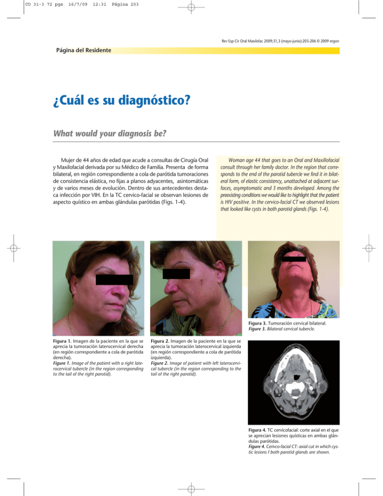
CO 31-3 72 pgs 16/7/09 12:31 Página 203 Rev Esp Cir Oral Maxilofac 2009;31,3 (mayo-junio):203-206 © 2009 ergon Página del Residente ¿Cuál es su diagnóstico? What would your diagnosis be? Mujer de 44 años de edad que acude a consultas de Cirugía Oral y Maxilofacial derivada por su Médico de Familia. Presenta de forma bilateral, en región correspondiente a cola de parótida tumoraciones de consistencia elástica, no fijas a planos adyacentes, asintomáticas y de varios meses de evolución. Dentro de sus antecedentes destaca infección por VIH. En la TC cervico-facial se observan lesiones de aspecto quístico en ambas glándulas parótidas (Figs. 1-4). Woman age 44 that goes to an Oral and Maxillofacial consult through her family doctor. In the region that corresponds to the end of the parotid tubercle we find it in bilateral form, of elastic consistency, unattached at adjacent surfaces, asymptomatic and 3 months developed. Among the preexisting conditions we would like to highlight that the patient is HIV positive. In the cervico-facial CT we observed lesions that looked like cysts in both parotid glands (Figs. 1-4). Figura 3. Tumoración cervical bilateral. Figure 3. Bilateral cervical tubercle. Figura 1. Imagen de la paciente en la que se aprecia la tumoración laterocervical derecha (en región correspondiente a cola de parótida derecha). Figure 1. Image of the patient with a right laterocervical tubercle (in the region corresponding to the tail of the right parotid). Figura 2. Imagen de la paciente en la que se aprecia la tumoración laterocervical izquierda (en región correspondiente a cola de parótida izquierda). Figure 2. Image of patient with left laterocervical tubercle (in the region corresponding to the tail of the right parotid). Figura 4. TC cervicofacial: corte axial en el que se aprecian lesiones quísticas en ambas glándulas parótidas. Figure 4. Cerivco-facial CT: axial cut in which cystic lesions f both parotid glands are shown. CO 31-3 72 pgs 16/7/09 12:31 Página 204 Rev Esp Cir Oral Maxilofac 2009;31,3 (mayo-junio):203-206 © 2009 ergon Página del Residente Lesión linfoepitelial quística benigna de glándula parótida en paciente con infección por VIH Lesion of benign lymphoepithileal cyst of the parotid gland in a patient infected with HIV C. Moreno García1, M.A. Pons García2, R. González García2, L. Ruiz Laza2, F. Monje Gil3 Introducción Introduction La Lesión Linfoepitelial Quística Benigna (LLQB) es un trastorno poco frecuente que afecta a las glándulas salivales, fundamentalmente a la glándula parótida y que se ha asociado a la infección por el VIH. Desde el punto de vista clínico, a nivel parotídeo y cervical, se manifiesta por la presencia de una tumoración elástica no dolorosa, de crecimiento progresivo y asociada a adenopatías cervicales. La LLQB es una entidad a tener en cuenta en el diagnóstico diferencial de las masas cervicales en pacientes infectados por el VIH. Lesion of Benign Lymphoepithelial Cyst (LBLC) is an uncommon condition that affects the salivary glands, mainly the parotid gland, and has been associated with HIV infection. From a clinical perspective, regarding parotid and cervical, they form because of the presence of an unpainful elastic tubercle growing progressively and associated with the cervical adenopathy. The LBLC is something to take into account during the differential diagnosis of cervical masses in patients infected with the HIV virus. Discusión Discussion La infección por el VIH se ha asociado a diversas entidades que afectan a las glándulas salivales como son el linfoma, síndrome de Sjögren, sarcoma de Kaposi y la lesión linfoepitelial quística benigna.1-6 Antes de la década de los ochenta las LLQB de parótida constituían menos del 3% de los tumores benignos de esta glándula, posteriormente y debido al aumento de la incidencia de la infección VIH, se ha incrementado el numero de casos.1,2 La LLQB de las glándulas salivales afecta con más frecuencia a la parótida que a las submaxilares.6-8 Histológicamente la lesión está compuesta por uno o varios quistes llenos de líquido gelatinoso claro, tapizados por epitelio metaplásico escamoso o columnar y rodeados por un infiltrado linfoide, que contiene islotes de células 1 Médico Residente. 2 Médico Adjunto. 3 Jefe de Servicio. Servicio de Cirugía Oral y Maxilofacial. Hospital Infanta Cristina. Complejo Hospitalario Universitario de Badajoz. España. Correspondencia: Carlos Moreno García Hospital Infanta Cristina Avenida de Elvas / Carretera de Portugal s/n 06080 Badajoz. España Email: carlosmorenogar@gmail.com Email: carlosmorenogarcia@wanadoo.es HIV infection is associated with many entities that affect the salivary glands, for example lymphoma, Sjögren syndrome, Kaposi sarcoma and lesion of benign lymphoepithelial cyst.1-6 Before the 80’s LBLC’s of the parotid made up less than 3% of the benign tumors reported in this gland. Later and due to the increased incidence of HIV infection the number of such cases increased.1, 2 LBLC of the salivary glands affects the parotid more frequently than the sub maxillas6-8. Histologically the lesion is made up of one or various cysts filled with a clear gelatinlike liquid, covered by a scaly metaplastic epithelium or columnar, and surrounded by an infiltrated lymphoid that has myoepithelial islet cells. The lymphoid component has the same characteristics as those seen in the adenopathys of the less advanced HIV infection, that is to say hyperplasia or follicle fragmentation with enlarged germinating centers. The etiology of the LBLC is unknown; indicators of active replication of HIV-1 like the p24 protein or viral RNA, in the sinus of reticular dendrictic cells of lymphoid follicles. Their histology is similar to that of the adenopathys of persistent poly adenopathic syndrome. As a result of this knowledge it occurred to some authors that these lesions are caused by HIV9. Another etiopathogenic hypothesis is based on the CO 31-3 72 pgs 16/7/09 12:31 Página 205 C. Moreno y cols. mioepiteliales. El componente linfoide tiene las mismas características que se observan en las adenopatías de la infección por VIH menos avanzada, es decir, hiperplasia o fragmentación folicular, con agrandamiento de los centros germinales.7,9-12 El infiltrado linfoide está mayoritariamente formado por células B, aunque existen numerosos linfocitos T, principalmente CD89. La etiología de la LLQB es desconocida; se han encontrado marcadores de replicación activa del VIH-1, como son la proteína p24 o el ARN viral, en el seno de las células dendríticas reticulares de los folículos linfoides y su histología es similar a las adenopatías del síndrome poliadenopático persistente. Todo esto, ha hecho sugerir a algunos autores, que estas lesiones son inducidas directamente por el VIH9. Otra hipótesis etiopatogénica se basa en que en la capsula parotídea quedarían englobados varios ganglios linfáticos con restos de acinis glandulares en su interior; la hiperplasia reactiva del tejido linfático, secundaria a la infección por VIH, obstruiría el ducto de excreción de los restos glandulares, dando lugar a quistes de retención.11,12,24 La forma clínica habitual de presentación consiste en una tumoración cervical indolora de crecimiento lento, sin signos inflamatorios, con frecuencia bilateral, aunque asimétrica. Suele aparecer en pacientes con síndrome poliadenopático persistente, con un número de linfocitos CD4 poco o medianamente disminuido y relativo aumento de células CD8 en sangre.6-9,13,16-18,23,25 Dentro de los estudios complementarios a realizar destacamos la TC, RM y ecografía; donde apreciaremos múltiples quistes parotídeos bilaterales asociados a adenopatías cervicales. Según distintos autores11,12 las imágenes de TC y RM en pacientes VIH son patognomónicas, y no requerirían más estudios complementarios. En cualquier caso podríamos completar el diagnostico mediante PAAF. Las manifestaciones clínicas responden habitualmente al tratamiento con antirretrovirales.11,19 Se han descrito casos en que la LLQB ha remitido al administrar tratamiento con zidovudina, aunque este hecho no ha sido constante.7 En ausencia de respuesta, y si el paciente refiere molestias locales o motivos estéticos se ha propuesto como tratamiento el drenaje percutáneo periódico de los quistes o la exéresis quirúrgica,11 (normalmente mediante parotidectomía superficial). El tratamiento mediante radioterapia a bajas dosis añade morbilidad a una patología benigna (xerostomía, inducción de lesiones neoplásicas, etc.) sobre todo en el caso de población pediátrica y los resultados obtenidos con dicho tratamiento han sido parciales.22 Como tratamiento minimamente invasivo se ha planteado la escleroterapia con doxiciclina, pero las series en las que se ha realizado son pequeñas y se precisa un seguimiento a más largo plazo de estos pacientes para comprobar resultados.21 Conclusiones La LLQB es una entidad que hemos de tener en cuenta en el diagnóstico diferencial de masas cervicales, en pacientes con infección por VIH. La benignidad de dicha lesión nos permitiría tratarla de forma conservadora, sobre todo en pacientes con inmunodepresión avanzada. Rev Esp Cir Oral Maxilofac 2009;31,3 (mayo-junio):203-206 © 2009 ergon 205 fact that the parotid capsule would remain lumped together in various lymphatic ganglion nodes with parts of acinic glands on the inside. The hyperplasia reactivates the lymphatic tissue, secondary to the HIV infection, obstructing the excretion duct of the remaining glands, allowing for cystic retention. The usual appearance consists of a slow growing unpainful cervical tubercle, without signs of inflammation, with bilateral symmetry. It usually occurs in patients with persistent polyadenopathic syndrome, with a slight or fair decrease in CD4 lymphocytes and a relative increase in CD8 cells in the blood.6-9,13,16-8,23,25 Within the complimentary studies when performing the CT, MRI and ecograph we find multiple bilateral parotid cysts associated with cervical adenopathys. According to different doctors the CT and MRI images of HIV patients are patognomonical and don’t need more complimentary studies. In any case we could finish the diagnosis using FNAB. Clinical manifestations usually respond to treatment with anti-retrovirals.11, 19 Many cases have been described where LBLC was treated with Zidovudine, although it wasn’t done constantly.7 In the absence of an answer if the patients reports local discomfort r esthetic motives periodic percutaneous drainage is the suggested treatment of these cysts. Another option is surgical abscission (normally using superficial parotidectomy). Treatment using low doses of radiotherapy adds morbidity to the benign pathology especially in pediatric cases. The results with this treatment have been partial.22 As a minimally invasive treatment sclerotherapy with doxycycline has been suggested, but the series that were carried out are small and long term follow up with these patients is necessary in order to compare results.21 Conclusions The LBLC is a topic that should be taken into account when diagnosing cervical masses found in HIV patients. This lesion’s benignancy allows us to treat it in a conservative manner especially in patients with advanced immunodepression. CO 31-3 72 pgs 206 16/7/09 12:31 Página 206 Rev Esp Cir Oral Maxilofac 2009;31,3 (mayo-junio):203-206 © 2009 ergon Bibliografía 1. Calderón E, Vinuesa M, Fernández P, Mendoza E, Gallardo JA, Pineda JA. Lesión linfoepitelial quística de parótida en pacientes infectados por el VIH. Medicina Clínica 1995;105:461-3. 2. Schiodt M, Dodd CL, Greenspan D, Daniels TE, Chernoff D, Hollander H et al. Natural history of HIV-associated salivary gland disease. Oral Surg Oral Med Oral Pathol 1992;74:326-31. 3. Mamary Y, Gomori JM, Nitzan DW. Lymphoepitelial parotid cysts as presenting symptom of immunodeficiency virus infection: clinical, sialographic, and magnetic resonance imaging findings. J Oral Maxillofac Surg 1990;48:981-4. 4. Ryan JR, Ioachim HL, Marmer J, Loubeau JM. Acquired immune deficiency syndrome-related lymphadenopathies presenting in the salivary gland lymph nodes. Arch Otolaryngol 1985;111:554-6. 5. Itescu S, Dalton J, Zhand HZ, Winchester R. Tissue infiltration in a CD8 lymphocytosis syndrome associated with human immunodeficiency virus-1 infection has the phenotypic appearance of anantigenically driven response. J Clin Invest 1993;91:2.216-2.225. 6. Terry JH, Loree TR, Thomas MD, Marti JR. Major salivary gland lymphoepithelial lesions andthe acquired immunodeficiency syndrome. Am J Surg 1991;162: 324-9. 7. Shaha AR, Di Maio T, Webber C, Thelmo W, Jaffe BM. Bening lymphoepithelial lesions of the parotid. Am J Surg 1993;166:403-7. 8. No-Louis E, Morales C, Jurado R, López-Beltrán A. Quiste linfoepitelial parotídeo e infección por virus de la inmunodeficiencia humana: diagnóstico citológico por punción aspirativa con aguja fina. Patología 1992;25:237-40. 9. Labouyrie E, Merlio JPH, Beylot-Barry M, DelordB, Vergier B, Brossard G et al. Human immunodeficiency virus type 1 replication within cystic lymphoepithelial lesion of the salivary gland. AmJ Clin Pathol 1993;100:41-6. 10. Flores JM, Andrada E, Pineda JA, Borderas F, Díaz MA, Soto B et al. Significado de la histología ganglionar en los pacientes con infección por el virus de inmunodeficiencia humana. Rev Clin Esp 1988;183:170-4. 11. Soler F, Concejo C, Borja A. Trastornos funcionales e inflamatorios de las glándulas salivares. Tratado de Cirugía Oral y Maxilofacial 2004;55:937. 12. Goto TK, Shimizu M, Kobayashi I, et al. Lymphoepitelial lesion of the parotid gland. Dentomaxillofac Radiol 2002;31:198-203. Lesión linfoepitelial quística benigna de glándula parótida en paciente con infección por vih 13. Fortuño-Mar A, Mayayo E, Castillo A, Guiral H. A Lymphoepitelial cyst of the parotid gland in an advanced stage of HIV infection. A rare association. An Otorrinolaringol Ibero Am 1999;2:469-75. 14. Rivas Lacarte MP, Javaloyas de Morlius M, Jiménez Fàbrega X, Oliván M, Torramilans A, Catalá Costa I. Intraparotid lymphoepithelial cysts and the human immunodeficiency virus. Rev Clin Esp 1997;197: 417-9. 15. Mandel L, Reich R. HIV parotid gland lymphoepithelial cysts. Review and case reports. Oral Surg Oral Med Oral Pathol 1992;74:273-8. 16. Favia G, Capodiferro S, Scivetti M, Lacaita MG, Filosa A, Lo Muzio L. Multiple parotid lymphoepithelial cysts in patients with HIV-infection: report of two cases. Oral Dis 2004;10:151-4. 17. Rahman S, Shaari R, Hassan R. Parotid Lymphoepithelial Cyst: A Case Report. Arch Orofac Scienc 2006;1:71-5. 18. Carrillo J, Urquijo ME, Hernández D, Vázquez C, Larrazar O. Quistes linfoepiteliales múltiples de la glándula parótida. Un padecimiento concomitante con infección por el VIH. Med Int Mex 2003;19:249-51. 19. Craven D, Duncan R, Stram J, O'Hara C, Steger K, Jhamb K, Hirschhorn L. Response of Lymphoepithelial Parotid Cysts to Antiretroviral Treatment in HIV-Infected Adults. Ann Internal Med 1998;128:4559. 20. Takahashi K, Uzawa N, Kosaka S, Yoshino N, Okada N, DDS, Amagasa T, DDS. Synchronous Warthin Tumors and Lymphoepithelial Cyst in the Ipsilateral Parotid Gland. J Oral Maxillofac Surg 2008;66:10536. 21. Suskind D, Tavill M, Handler S. Doxycycline sclerotherapy of benign lymphoepithelial cysts of the parotid: a minimally invasive treatment. Int J Ped Otorhinolaryngol 2000;52:157-61. 22. Goldstein J, Rubin J, Silver C. Radiation therapy as a treatment for benign lymphoepithelial parotid cysts inpatients infected with human immunodeficiency virus-1, Int J Radiol Oncol Biol Phys 1992;23:104550. 23. Dave SP, Pernas FG, Roy S. The benign lymphoepithelial cyst and a classification system for lymphocytic parotid gland enlargement in the pediatric HIV population. Laryngoscope 2007;117:106-13. 24. Tao LC, Gullane P. HIV infection-associated lymphoepithelial lesions of the parotid gland: Aspiration biopsy cytology, histology, and pathogenesis. Diagnostic Cytopathology 2006;7:158-62. 25. Zeitlen S, Shaha A. Parotid manifestations of HIV infection. J Surg Oncol 2006;47:230-2.
