Quantitative Analysis of the Mobility of the Left Ventricle Using
Anuncio
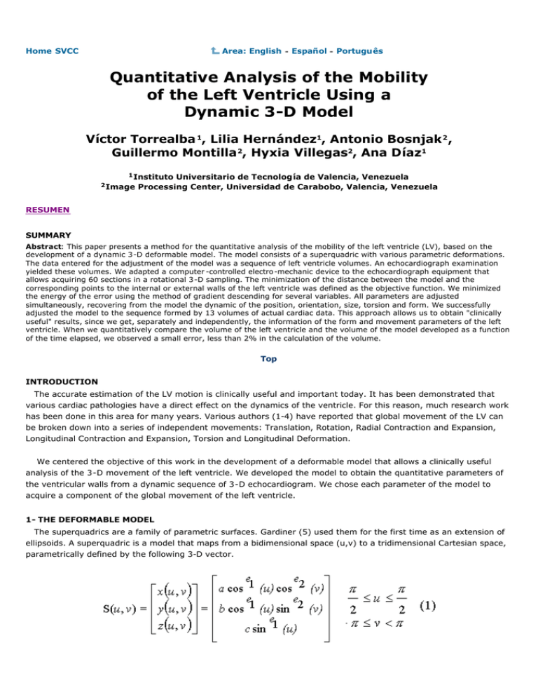
Home SVCC Area: English - Español - Português Quantitative Analysis of the Mobility of the Left Ventricle Using a Dynamic 3-D Model Víctor Torrealba 1, Lilia Hernández1, Antonio Bosnjak 2 , Guillermo Montilla 2, Hyxia Villegas2, Ana Díaz1 1 Instituto 2 Image Universitario de Tecnología de Valencia, Venezuela Processing Center, Universidad de Carabobo, Valencia, Venezuela RESUMEN SUMMARY Abstract: This paper presents a method for the quantitative analysis of the mobility of the left ventricle (LV), based on the development of a dynamic 3-D deformable model. The model consists of a superquadric with various parametric deformations. The data entered for the adjustment of the model was a sequence of left ventricle volumes. An echocardiograph examination yielded these volumes. We adapted a computer -controlled electro-mechanic device to the echocardiograph equipment that allows acquiring 60 sections in a rotational 3-D sampling. The minimization of the distance between the model and the corresponding points to the internal or external walls of the left ventricle was defined as the objective function. We minimized the energy of the error using the method of gradient descending for several variables. All parameters are adjusted simultaneously, recovering from the model the dynamic of the position, orientation, size, torsion and form. We successfully adjusted the model to the sequence formed by 13 volumes of actual cardiac data. This approach allows us to obtain "clinically useful" results, since we get, separately and independently, the information of the form and movement parameters of the left ventricle. When we quantitatively compare the volume of the left ventricle and the volume of the model developed as a function of the time elapsed, we observed a small error, less than 2% in the calculation of the volume. Top INTRODUCTION The accurate estimation of the LV motion is clinically useful and important today. It has been demonstrated that various cardiac pathologies have a direct effect on the dynamics of the ventricle. For this reason, much research work has been done in this area for many years. Various authors (1-4) have reported that global movement of the LV can be broken down into a series of independent movements: Translation, Rotation, Radial Contraction and Expansion, Longitudinal Contraction and Expansion, Torsion and Longitudinal Deformation. We centered the objective of this work in the development of a deformable model that allows a clinically useful analysis of the 3-D movement of the left ventricle. We developed the model to obtain the quantitative parameters of the ventricular walls from a dynamic sequence of 3-D echocardiogram. We chose each parameter of the model to acquire a component of the global movement of the left ventricle. 1- THE DEFORMABLE MODEL The superquadrics are a family of parametric surfaces. Gardiner (5) used them for the first time as an extension of ellipsoids. A superquadric is a model that maps from a bidimensional space (u,v) to a tridimensional Cartesian space, parametrically defined by the following 3-D vector. The u and v parameters correspond, respectively, to the latitude and longitude angles expressed in a system of spherical coordinates centered on the object. The scale parameters a, b and c define the size of the superquadric in the x, y, and z coordinates, respectively. The exponents e 1 and e 2 produce an squareness effect of the ellipsoid. Varying the size of the three semi-axes forming the superquadric (a, b and c), modifies its global size ( Figure 1 ). The equation 1 define the model according to five parameters that shapes it, within a system of coordinates centered in the model. To move the model in order to fit it to the surface of the left ventricle, we defined an homogeneous transformation matrix T, expressed in the following manner: The Euler angles (φ, θ , ψ ) are used to express the rotational part of the transform. The values px, p y and p z indicate the translation of the model's coordinate system. The transform of equation (2) adds 6 parameters to the model to make 11 parameters a, b, c, e1, e 2, φ, θ , ψ , p x, p y , p z. They allow the generation of a superquadric centered in an arbitrary spatial location and an arbitrary orientation of its principal axes. The Tapering and Torsion deformations add two additional transforms performed on the model. The Tapering changes the size of the superquadric along the axes x, y, according to the spatial location with regards to the axis z. Figure 2 shows the effect of this transform. The following equation defines the Tapering deformation: By adding the Torsion deformation ( Figure 3) to the model we obtain the torsion movement proper of the left ventricle and define it by the following transform: The Tapering and Torsion transforms add two additional parameters to the model, creating a model with thirteen parameters. 2- ADJUSTMENT OF THE MODEL 3.1 Data used To verify the model's behavior, we use a 3-D sequence of actual cardiac data made up of thirteen volumes acquired in a cardiac period. The authors obtained this data in previous works (6). To acquire this sequence we used the method of transthoracic rotational sweep and for each time interval we make sixty (60) radial sections of the ventricle. and we finally extract the edges of the ventricle's internal and external walls. We store the spatial data of the position of each of the points forming the edges of the ventricular walls as vectors and this cloud of points is used for the subsequent adjustment of the model. 3.2 Adjustment of the Model Parameters We minimized the energy of the error for the adjustment of the model's parameters using the method of gradient descending for several variables. We defined as objective function to minimize the following error expression between the model and the data: where: pn: are the points obtained from the segmentation of the ventricle. ai : are the parameters of the model. F(pn:ai ) is the implicit equation of the superquadric with all transforms, with 13 parameters The F(pn:ai ) function takes the following values: The gradient vector of the error function indicates the direction of growth of the objective function. Therefore, the ai parameters are modified in small increases in the opposite direction. We performed the iterative adjustment until the variation of each of the ai parameters is less than an e value previously established. 3.4 RESULTS When the model's adjustment is completed, the position, orientation, size, torsion and shape dynamics of the left ventricle are recovered. The model adjusts to all the sequence of volumes of actual cardiac data. The results obtained are the information of the dynamics for the translation, orientation, radial and longitudinal contraction/expansion (Figure 4 ), torsion ( Figure 5 ), and the longitudinal deformation parameters of the model. Figure 6 simultaneously shows the left ventricle and the model developed in the instants of telediastole and telesystole. To quantitatively compare the model developed against the left ventricle, the volume of the ventricular cavity is graphed as a function of time ( Figure 7). Figure 8 shows the percentage error made using this model as a function of time elapsed. Another measure of comparison between the model and left ventricle was the ejection fraction that generated the following results: Real ejection fraction 52.79% Model ejection fraction 52.10% 3.4 CONCLUSIONS We presented in this work a parametric model for the quantitative analysis of the 3-D analysis of the movement of the left ventricle. The model developed furnishes the means to obtain "clinically useful" results, because the left ventricle's translation, rotation, longitudinal contraction, expansion, and the longitudinal deformation data are obtained separately and independently. The manner of quantitatively presenting the results of the movement dynamics leads us to suppose that the results obtained will be easily interpreted by the medical specialists. If we use the data obtained by means of the model and we evaluate it against a 3-D echocardiograph database with sufficient normal and pathological cases, it is possible that this small group of parameters will be enough to distinguish between a normal and a pathological case. Thus, it helps to guide the patient's diagnosis and the therapy to follow in each particular case. An additional contribution of the work is the adjustment of the model's parameters. It showed its robustness by automatically recovering 26 superquadrics (internal and external ventricle walls) and its deformations from a sequence of original volumes. The next step is developing an automatic 3-D segmentation method of the ventricular walls, based on the model developed. Top Modelo 3D con Deformaciones Paramétricas para el An álisis de la Movilidad 3D del Ventrículo Izquierdo RESUMEN Introducci ón: La medida del movimiento de las paredes del ventr ículo izquierdo de una manera exacta es muy importante en la actualidad, ya que se ha demostrado que diferentes patolog ías cardiacas tienen directa repercusión sobre la dinámica del ventrículo. Diferentes autores han reportado que durante el ciclo cardiaco el movimiento global del ventrículo izquierdo se puede descomponer en una serie de movimientos independientes, los cuales son: Traslación, Rotación, Contracción y Expansión Radial, Torsi ón, Contracción y Expansión Longitudinal, Deformación Longitudinal. Objetivos: El objetivo de este trabajo se centr ó en el desarrollo de un modelo deformable que permite cuantificar, graficar y por consiguiente analizar la dinámica del movimiento 3D del ventrículo izquierdo. Material y Métodos : El modelo consiste en una supercuádrica con diferentes deformaciones paramétricas, la cual se ajusto a una secuencia de 13 volúmenes del ventr ículo izquierdo previamente segmentados por un cardiologo especialista. Se definió como función objetivo para el ajuste de los parámetros del modelo la minimización de la distancia de todos los puntos del ventrículo izquierdo al modelo desarrollado. Los volúmenes fueron obtenidos por medio de una estación de captura, visualización y reconstrucción de data cardiaca 3D desarrollada por los autores en trabajos previos. Resultados: El enfoque con que se orientó el desarrollo del modelo, permite obtener resultados "clínicamente útiles" ya que se obtienen por separado y de forma independiente la información de los movimientos de traslación, rotación, contracción y expansión radial, torsión, contracción y expansi ón longitudinal y deformaci ón longitudinal del ventrículo izquierdo. Discusión: La forma sencilla de presentar cuantitativamente los resultados de la dinámica del movimiento (en forma similar a las graficas de ECG), hace suponer que los resultados obtenidos serán fácilmente interpretados por los médicos especialistas. Conclusiones: Es de suponer que si en un futuro se dispone de una base de datos con suficientes casos normales y patológicos, a partir del an álisis de la información capturada por el modelo, se podrán diagnosticar fácilmente diferentes patologías cardíacas. REFERENCES 1. J. Park, "Analysis of the ventricular wall motion based on volumetric deformable models and MRI -SPAMM", Medical Image Analysis, 1, 53-71 (1998). 2. A. Hunter, "Description of the deformation of the ventricle by a kinematic model" Journal Biomech, 243, 1119-1127 (1994). 3. J. Puentes, "Analyze symbolique du mouvement cardiaque en Angiographie Vasculaire". Tesis Doctoral (1998). 4. M Marcus, "Determinants of systolic and diastolic Ventricular Function", Cardiac Imaging (1991). 5. M. Gardiner, "The superellipse: A curve that lies betwen the ellipse and the rectangle", Sci. Amer, 213, 222-234 (1965). 6. V. Torrealba, "Ecocardiografía Dinámica Tridimensional. Estación de Trabajo para la Adquisición, Reconstrucción y Visualización de Imágenes 4D del Corazón" http://www.ciudadfutura.com/ecocardiografia/trabajos/estacion4d/ecocardi.htm (2000.) 7. Chong-Huah, "3D Moments Forms: Their Construction and Application to Object Identification and Positioning" IEEE-PAMI, 11 (1991). Top Your questions, contributions and commentaries will be answered by the authors in the Echocardiography list. Please fill in the form (in Spanish, Portuguese or English) and press the "Send" button. Question, contribution or commentary: Name and Surname: Country: Argentina E-Mail address: @ Send Erase Top 2nd Virtual Congress of Cardiology Dr. Florencio Garófalo Dr. Raúl Bretal Dr. Armando Pacher Steering Committee President Scientific Committee President Technical Committee - CETIFAC President fgaro@fac.org.ar fgaro@satlink.com rbretal@fac.org.ar rbretal@netverk.com.ar apacher@fac.org.ar apacher@satlink.com Copyright© 1999-2001 Argentine Federation of Cardiology All rights reserved This company contributed to the Congress:
