Anuncio
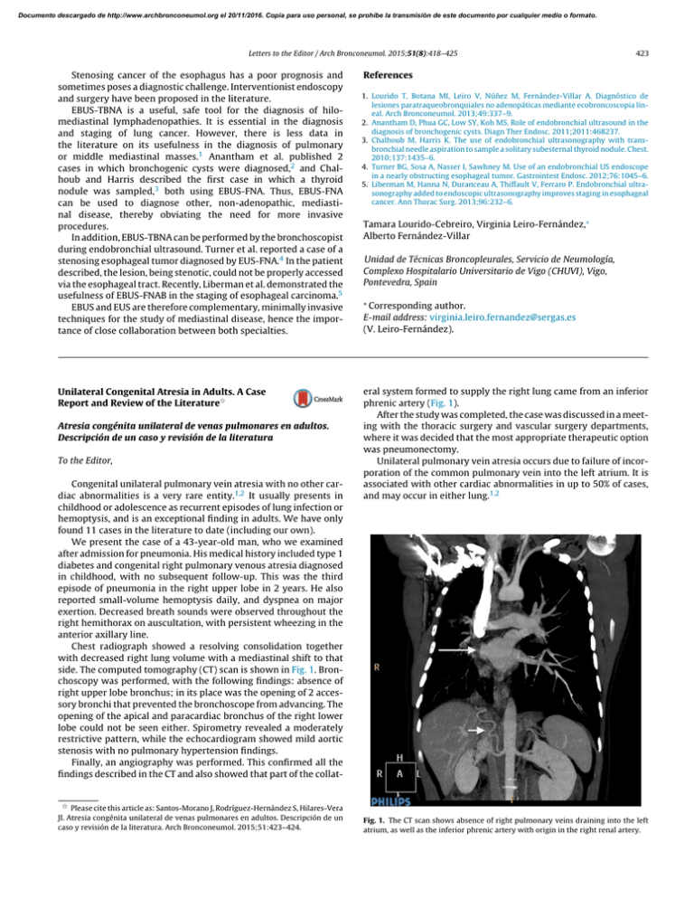
Documento descargado de http://www.archbronconeumol.org el 20/11/2016. Copia para uso personal, se prohíbe la transmisión de este documento por cualquier medio o formato. Letters to the Editor / Arch Bronconeumol. 2015;51(8):418–425 Stenosing cancer of the esophagus has a poor prognosis and sometimes poses a diagnostic challenge. Interventionist endoscopy and surgery have been proposed in the literature. EBUS-TBNA is a useful, safe tool for the diagnosis of hilomediastinal lymphadenopathies. It is essential in the diagnosis and staging of lung cancer. However, there is less data in the literature on its usefulness in the diagnosis of pulmonary or middle mediastinal masses.1 Anantham et al. published 2 cases in which bronchogenic cysts were diagnosed,2 and Chalhoub and Harris described the first case in which a thyroid nodule was sampled,3 both using EBUS-FNA. Thus, EBUS-FNA can be used to diagnose other, non-adenopathic, mediastinal disease, thereby obviating the need for more invasive procedures. In addition, EBUS-TBNA can be performed by the bronchoscopist during endobronchial ultrasound. Turner et al. reported a case of a stenosing esophageal tumor diagnosed by EUS-FNA.4 In the patient described, the lesion, being stenotic, could not be properly accessed via the esophageal tract. Recently, Liberman et al. demonstrated the usefulness of EBUS-FNAB in the staging of esophageal carcinoma.5 EBUS and EUS are therefore complementary, minimally invasive techniques for the study of mediastinal disease, hence the importance of close collaboration between both specialties. Unilateral Congenital Atresia in Adults. A Case Report and Review of the Literature夽 Atresia congénita unilateral de venas pulmonares en adultos. Descripción de un caso y revisión de la literatura To the Editor, Congenital unilateral pulmonary vein atresia with no other cardiac abnormalities is a very rare entity.1,2 It usually presents in childhood or adolescence as recurrent episodes of lung infection or hemoptysis, and is an exceptional finding in adults. We have only found 11 cases in the literature to date (including our own). We present the case of a 43-year-old man, who we examined after admission for pneumonia. His medical history included type 1 diabetes and congenital right pulmonary venous atresia diagnosed in childhood, with no subsequent follow-up. This was the third episode of pneumonia in the right upper lobe in 2 years. He also reported small-volume hemoptysis daily, and dyspnea on major exertion. Decreased breath sounds were observed throughout the right hemithorax on auscultation, with persistent wheezing in the anterior axillary line. Chest radiograph showed a resolving consolidation together with decreased right lung volume with a mediastinal shift to that side. The computed tomography (CT) scan is shown in Fig. 1. Bronchoscopy was performed, with the following findings: absence of right upper lobe bronchus; in its place was the opening of 2 accessory bronchi that prevented the bronchoscope from advancing. The opening of the apical and paracardiac bronchus of the right lower lobe could not be seen either. Spirometry revealed a moderately restrictive pattern, while the echocardiogram showed mild aortic stenosis with no pulmonary hypertension findings. Finally, an angiography was performed. This confirmed all the findings described in the CT and also showed that part of the collat- 夽 Please cite this article as: Santos-Morano J, Rodríguez-Hernández S, Hilares-Vera JI. Atresia congénita unilateral de venas pulmonares en adultos. Descripción de un caso y revisión de la literatura. Arch Bronconeumol. 2015;51:423–424. 423 References 1. Lourido T, Botana MI, Leiro V, Núñez M, Fernández-Villar A. Diagnóstico de lesiones paratraqueobronquiales no adenopáticas mediante ecobroncoscopia lineal. Arch Bronconeumol. 2013;49:337–9. 2. Anantham D, Phua GC, Low SY, Koh MS. Role of endobronchial ultrasound in the diagnosis of bronchogenic cysts. Diagn Ther Endosc. 2011;2011:468237. 3. Chalhoub M, Harris K. The use of endobronchial ultrasonography with transbronchial needle aspiration to sample a solitary subesternal thyroid nodule. Chest. 2010;137:1435–6. 4. Turner BG, Sosa A, Nasser I, Sawhney M. Use of an endobronchial US endoscope in a nearly obstructing esophageal tumor. Gastrointest Endosc. 2012;76:1045–6. 5. Liberman M, Hanna N, Duranceau A, Thiffault V, Ferraro P. Endobronchial ultrasonography added to endoscopic ultrasonography improves staging in esophageal cancer. Ann Thorac Surg. 2013;96:232–6. Tamara Lourido-Cebreiro, Virginia Leiro-Fernández,∗ Alberto Fernández-Villar Unidad de Técnicas Broncopleurales, Servicio de Neumología, Complexo Hospitalario Universitario de Vigo (CHUVI), Vigo, Pontevedra, Spain ∗ Corresponding author. E-mail address: virginia.leiro.fernandez@sergas.es (V. Leiro-Fernández). eral system formed to supply the right lung came from an inferior phrenic artery (Fig. 1). After the study was completed, the case was discussed in a meeting with the thoracic surgery and vascular surgery departments, where it was decided that the most appropriate therapeutic option was pneumonectomy. Unilateral pulmonary vein atresia occurs due to failure of incorporation of the common pulmonary vein into the left atrium. It is associated with other cardiac abnormalities in up to 50% of cases, and may occur in either lung.1,2 Fig. 1. The CT scan shows absence of right pulmonary veins draining into the left atrium, as well as the inferior phrenic artery with origin in the right renal artery. Documento descargado de http://www.archbronconeumol.org el 20/11/2016. Copia para uso personal, se prohíbe la transmisión de este documento por cualquier medio o formato. 424 Letters to the Editor / Arch Bronconeumol. 2015;51(8):418–425 The clinical spectrum ranges from asymptomatic patients3 to the development of pulmonary hypertension2 and death. Diagnosis is confirmed by pulmonary angiography. However, modern techniques such as CT scan or magnetic resonance imaging together with symptoms consistent with the condition may now be sufficient to make the diagnosis.4 There are 3 therapeutic options proposed in the literature. Pneumonectomy has been shown to control symptoms, and to prevent pulmonary hypertension.1,5 Another, less aggressive, therapeutic option is coil embolization of the systemic arterial collateral vessels. In the article by Heyneman et al.,2 this procedure controlled hemoptysis in one of the patients, who subsequently remained asymptomatic. The third option, in cases of asymptomatic patients or those with few symptoms, is follow-up, focusing particularly on the early detection of pulmonary hypertension. 2. Heyneman LE, Nolan RL, Harrison JK, McAdams HP. Congenital unilateral pulmonary vein atresia: radiologic findings in three adult patients. Am J Roentgenol. 2001;177:681–5. 3. Kim Y, Yoo IR, Ahn MI, Han DH. Asymptomatic adults with isolated, unilateral right pulmonary vein atresia: multidetector CT findings. Br J Radiol. 2011;84: 109–13. 4. Artero Muñoz I, Serrano Puche F, Padín Marín MI, Serrano Ramos F. Congenital unilateral pulmonary vein atresia: imaging findings. Radiologia. 2008;50:82–5 [Article in Spanish]. 5. Swischuk LE, L’Heureux P. Unilateral pulmonary vein atresia. Am J Roentgenol. 1980;135:667–72. Juan Santos-Morano,a,∗ Silvia Rodríguez-Hernández,b Jesica Ivana Hilares-Veraa a References Servicio de Neumología, Hospital de Valme, Sevilla, Spain Servicio de Cuidados Críticos y Urgencias, Hospital de Valme, Sevilla, Spain 1. Pourmoghadam KK, Moore JW, Khan M, Geary EM, Madan N, Wolfson BJ, et al. Congenital unilateral pulmonary venous atresia: definitive diagnosis and treatment. Pediatr Cardiol. 2003;24:73–9. ∗ Corresponding author. E-mail address: juansan81@hotmail.com (J. Santos-Morano). Isolated Unilateral Pulmonary Vein Atresia in Adults夽 right hilum. The right pulmonary veins could not be identified on computed tomography (CT), and a diffuse increase in density was observed in the affected lung. Cardiac magnetic resonance imaging (MRI) ruled out associated heart disease, and the echocardiography ruled out pulmonary hypertension. Pulmonary arteriography was performed (Fig. 1A and B), in which hypoplasia of the right pulmonary artery and absence of right pulmonary venous return were observed. Arteriography of the thoracic artery (Fig. 1C) showed right bronchial artery hypertrophy originating in the descending aorta. Lung function test results were: FEV1: 2.040 (75.5%), FVC: 2.800 (90.1%) and DLCOc SB: 69.3%. Varices were observed in the distal trachea and start of the right main bronchus on fiber optic bronchoscopy. Considering the previous findings, we opted for surgical treatment, specifically, right pneumonectomy. The immediate post-operative period was uneventful, and the patient is currently asymptomatic. b Atresia aislada unilateral de venas pulmonares en el adulto To the Editor, We present the case of a 19-year-old woman, smoker (8 packyears), and regular cannabis user, with a history of severe bronchitis in childhood and frequent respiratory infections. She presented for episodes of coughing with hemoptoic expectoration since she was 15-years-old. The episodes were initially sporadic, but had become more frequent in recent months, sometimes occurring every 48 h. On examination, she had decreased breath sounds in the right hemithorax, and acropachy. The chest radiograph showed loss of volume in the right lung and attenuation of the Fig. 1. Pulmonary arteriography showing hypoplasia of the right pulmonary artery, providing little perfusion, almost entirely limited to the upper lobe (A), in which right venous drainage cannot be seen (B). Arteriography of the thoracic aorta showing an irregular, winding bronchial artery with origin in the anterior superior side of the descending thoracic aorta (C). 夽 Please cite this article as: Gómez Hernández MT, Rodríguez Pérez M, Jiménez MF. Atresia aislada unilateral de venas pulmonares en el adulto. Arch Bronconeumol. 2015;51:424–425.
