Unilateral Nevoid Hyperkeratosis of the Nipple and Areola
Anuncio
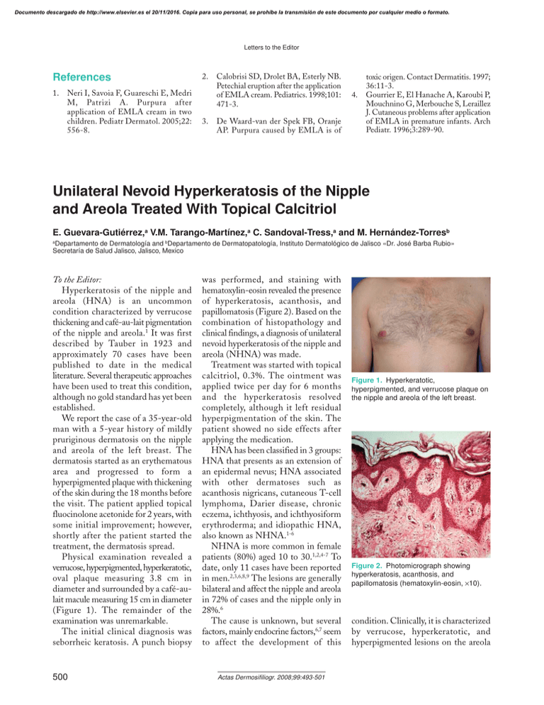
Documento descargado de http://www.elsevier.es el 20/11/2016. Copia para uso personal, se prohíbe la transmisión de este documento por cualquier medio o formato. Letters to the Editor References 1. Neri I, Savoia F, Guareschi E, Medri M, Patrizi A. Purpura after application of EMLA cream in two children. Pediatr Dermatol. 2005;22: 556-8. 2. 3. Calobrisi SD, Drolet BA, Esterly NB. Petechial eruption after the application of EMLA cream. Pediatrics. 1998;101: 471-3. De Waard-van der Spek FB, Oranje AP. Purpura caused by EMLA is of 4. toxic origen. Contact Dermatitis. 1997; 36:11-3. Gourrier E, El Hanache A, Karoubi P, Mouchnino G, Merbouche S, Leraillez J. Cutaneous problems after application of EMLA in premature infants. Arch Pediatr. 1996;3:289-90. Unilateral Nevoid Hyperkeratosis of the Nipple and Areola Treated With Topical Calcitriol E. Guevara-Gutiérrez,a V.M. Tarango-Martínez,a C. Sandoval-Tress,a and M. Hernández-Torresb a Departamento de Dermatología and bDepartamento de Dermatopatología, Instituto Dermatológico de Jalisco «Dr. José Barba Rubio» Secretaría de Salud Jalisco, Jalisco, Mexico To the Editor: Hyperkeratosis of the nipple and areola (HNA) is an uncommon condition characterized by verrucose thickening and café-au-lait pigmentation of the nipple and areola.1 It was first described by Tauber in 1923 and approximately 70 cases have been published to date in the medical literature. Several therapeutic approaches have been used to treat this condition, although no gold standard has yet been established. We report the case of a 35-year-old man with a 5-year history of mildly pruriginous dermatosis on the nipple and areola of the left breast. The dermatosis started as an erythematous area and progressed to form a hyperpigmented plaque with thickening of the skin during the 18 months before the visit. The patient applied topical fluocinolone acetonide for 2 years, with some initial improvement; however, shortly after the patient started the treatment, the dermatosis spread. Physical examination revealed a verrucose, hyperpigmented, hyperkeratotic, oval plaque measuring 3.8 cm in diameter and surrounded by a café-aulait macule measuring 15 cm in diameter (Figure 1). The remainder of the examination was unremarkable. The initial clinical diagnosis was seborrheic keratosis. A punch biopsy 500 was performed, and staining with hematoxylin-eosin revealed the presence of hyperkeratosis, acanthosis, and papillomatosis (Figure 2). Based on the combination of histopathology and clinical findings, a diagnosis of unilateral nevoid hyperkeratosis of the nipple and areola (NHNA) was made. Treatment was started with topical calcitriol, 0.3%. The ointment was applied twice per day for 6 months and the hyperkeratosis resolved completely, although it left residual hyperpigmentation of the skin. The patient showed no side effects after applying the medication. HNA has been classified in 3 groups: HNA that presents as an extension of an epidermal nevus; HNA associated with other dermatoses such as acanthosis nigricans, cutaneous T-cell lymphoma, Darier disease, chronic eczema, ichthyosis, and ichthyosiform erythroderma; and idiopathic HNA, also known as NHNA.1-6 NHNA is more common in female patients (80%) aged 10 to 30.1,2,4-7 To date, only 11 cases have been reported in men.2,3,6,8,9 The lesions are generally bilateral and affect the nipple and areola in 72% of cases and the nipple only in 28%.6 The cause is unknown, but several factors, mainly endocrine factors,6,7 seem to affect the development of this Actas Dermosifiliogr. 2008;99:493-501 Figure 1. Hyperkeratotic, hyperpigmented, and verrucose plaque on the nipple and areola of the left breast. Figure 2. Photomicrograph showing hyperkeratosis, acanthosis, and papillomatosis (hematoxylin-eosin, ×10). condition. Clinically, it is characterized by verrucose, hyperkeratotic, and hyperpigmented lesions on the areola Documento descargado de http://www.elsevier.es el 20/11/2016. Copia para uso personal, se prohíbe la transmisión de este documento por cualquier medio o formato. Letters to the Editor that extend to the nipples.3 The lesions are generally asymptomatic,5-8 although a case of NHNA reported by PérezIzquierdo et al6 prevented lactation. The typical histopathologic findings are hyperkeratosis with the formation of keratotic plugs, acanthosis, and papillomatosis.7 The differential clinical diagnoses to be considered are Paget disease, superficial basal cell carcinoma, dermatophytosis, and Bowen disease. The differential histopathologic diagnoses are verrucose epidermal nevus and acanthosis nigricans.1 Some therapeutic options that have proven effective in the treatment of this condition are salicylic acid, 6%, lactic acid lotion, topical corticosteroids, topical tretinoin, oral vitamin A, and topical calcipotriol.8,10 Kubota et al9 reported favorable cosmetic results with cryotherapy using liquid nitrogen. Surgical resection is also effective.1 This case is interesting, as NHNA is an uncommon condition, especially in men. Furthermore, although vitamin D analogues have traditionally been used in the past to treat this condition, the case presented here is the first in which topical calcitriol has been used. This agent inhibits cell proliferation and induces keratinocyte differentiation; therefore, it can be an option for therapy, although studies with larger numbers of patients are necessary. References 1. Mehregan AH, Rahbari H. Hyperkeratosis of nipple and areola. Arch Dermatol. 1977;113:1691-2. 2. Ahn SK, Chung J, Soo Lee W, Kim SC, Lee SH. Hyperkeratosis of the nipple and areola simultaneously developing with cutaneous T-cell lymphoma. J Am Acad Dermatol. 1995;32:124-5. 3. Allegue F, Soria C, Rocamora A, Fraile G, Ledo A. Hyperkeratosis of the nipple and areola in a patient with cutaneous T-cell lymphoma. Int J Dermatol. 1990;29:519-20. Actas Dermosifiliogr. 2008;99:493-501 4. Revert A, Bañuls J, Montesinos E, Jorda E, Ramon D, Torres V. Nevoid hyperkeratosis of the areola. Int J Dermatol. 1993;32:745-6. 5. Mehanna A, Malak JA, Kibbi AG. Hyperkeratosis of the nipple and areola: report of 3 cases. Arch Dermatol. 2001; 137:1327-8. 6. Pérez-Izquierdo JM, Vilata JJ, Sánchez JL, Gargallo E, Millan F, Aliaga A. Retinoic acid treatment of nipple hyperkeratosis. Arch Dermatol. 1990;126: 687-8. 7. D’Souza M, Gharami R, Ratnakar C, Garg BR. Unilateral nevoid hyperkeratosis of the nipple and areola. Int J Dermatol. 1996;35:602-3. 8. Bayramgurler D, Bilen N, Apaydin R, Ercin C. Nevoid hyperkeratosis of the nipple and areola: treatment of two patients with topical calcipotriol. J Am Acad Dermatol. 2002;46:131-3. 9. Kubota Y, Koga T, Nakayama J, Kiryu H. Naevoid hyperkeratosis of the nipple and areola in a man. Br J Dermatol. 2000;142:382-4. 10. Ruiz-Villaverde R, Sánchez-Cano D, Pacheco Sánchez Lafuente FJ. Hiperqueratosis nevoide del pezón y la areola. An Pediatr (Barc). 2006;64:180-1. 501
