Electrocardiographic Findings Typical of Brugada Syndrome
Anuncio
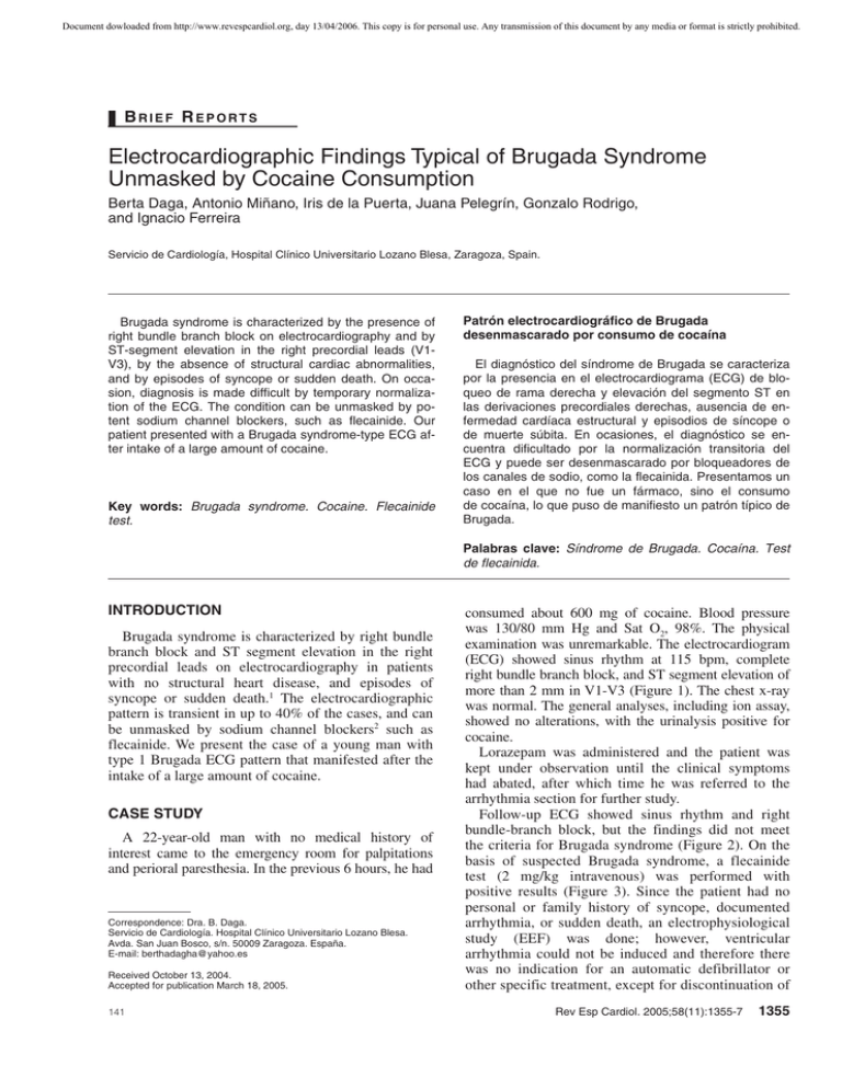
Document dowloaded from http://www.revespcardiol.org, day 13/04/2006. This copy is for personal use. Any transmission of this document by any media or format is strictly prohibited. BRIEF REPORTS Electrocardiographic Findings Typical of Brugada Syndrome Unmasked by Cocaine Consumption Berta Daga, Antonio Miñano, Iris de la Puerta, Juana Pelegrín, Gonzalo Rodrigo, and Ignacio Ferreira Servicio de Cardiología, Hospital Clínico Universitario Lozano Blesa, Zaragoza, Spain. Brugada syndrome is characterized by the presence of right bundle branch block on electrocardiography and by ST-segment elevation in the right precordial leads (V1V3), by the absence of structural cardiac abnormalities, and by episodes of syncope or sudden death. On occasion, diagnosis is made difficult by temporary normalization of the ECG. The condition can be unmasked by potent sodium channel blockers, such as flecainide. Our patient presented with a Brugada syndrome-type ECG after intake of a large amount of cocaine. Key words: Brugada syndrome. Cocaine. Flecainide test. Patrón electrocardiográfico de Brugada desenmascarado por consumo de cocaína El diagnóstico del síndrome de Brugada se caracteriza por la presencia en el electrocardiograma (ECG) de bloqueo de rama derecha y elevación del segmento ST en las derivaciones precordiales derechas, ausencia de enfermedad cardíaca estructural y episodios de síncope o de muerte súbita. En ocasiones, el diagnóstico se encuentra dificultado por la normalización transitoria del ECG y puede ser desenmascarado por bloqueadores de los canales de sodio, como la flecainida. Presentamos un caso en el que no fue un fármaco, sino el consumo de cocaína, lo que puso de manifiesto un patrón típico de Brugada. Palabras clave: Síndrome de Brugada. Cocaína. Test de flecainida. INTRODUCTION Brugada syndrome is characterized by right bundle branch block and ST segment elevation in the right precordial leads on electrocardiography in patients with no structural heart disease, and episodes of syncope or sudden death.1 The electrocardiographic pattern is transient in up to 40% of the cases, and can be unmasked by sodium channel blockers2 such as flecainide. We present the case of a young man with type 1 Brugada ECG pattern that manifested after the intake of a large amount of cocaine. CASE STUDY A 22-year-old man with no medical history of interest came to the emergency room for palpitations and perioral paresthesia. In the previous 6 hours, he had Correspondence: Dra. B. Daga. Servicio de Cardiología. Hospital Clínico Universitario Lozano Blesa. Avda. San Juan Bosco, s/n. 50009 Zaragoza. España. E-mail: berthadagha@yahoo.es Received October 13, 2004. Accepted for publication March 18, 2005. 141 consumed about 600 mg of cocaine. Blood pressure was 130/80 mm Hg and Sat O2, 98%. The physical examination was unremarkable. The electrocardiogram (ECG) showed sinus rhythm at 115 bpm, complete right bundle branch block, and ST segment elevation of more than 2 mm in V1-V3 (Figure 1). The chest x-ray was normal. The general analyses, including ion assay, showed no alterations, with the urinalysis positive for cocaine. Lorazepam was administered and the patient was kept under observation until the clinical symptoms had abated, after which time he was referred to the arrhythmia section for further study. Follow-up ECG showed sinus rhythm and right bundle-branch block, but the findings did not meet the criteria for Brugada syndrome (Figure 2). On the basis of suspected Brugada syndrome, a flecainide test (2 mg/kg intravenous) was performed with positive results (Figure 3). Since the patient had no personal or family history of syncope, documented arrhythmia, or sudden death, an electrophysiological study (EEF) was done; however, ventricular arrhythmia could not be induced and therefore there was no indication for an automatic defibrillator or other specific treatment, except for discontinuation of Rev Esp Cardiol. 2005;58(11):1355-7 1355 Document dowloaded from http://www.revespcardiol.org, day 13/04/2006. This copy is for personal use. Any transmission of this document by any media or format is strictly prohibited. Daga B, et al. Electrocardiographic Findings Typical of Brugada Syndrome Unmasked by Cocaine Consumption Figure 1. 12-lead electrocardiogram after cocaine consumption. Type 1 Brugada ECG pattern. Figure 2. 12-lead electrocardiogram at baseline. Figure 3. Electrocardiogram before administration of flecainide (A). Electrocardiogram after administration of flecainide. Positive pharmacological test (B). cocaine consumption. The patient required outpatient follow-up. In a subsequent study of near relatives, the father’s ECG showed right bundle branch block, but without the criteria defining a Brugada pattern. A flecainide test and EEF were also done, both with negative results. 1356 Rev Esp Cardiol. 2005;58(11):1355-7 DISCUSSION Brugada syndrome is a genetically determined disease caused by mutations in the sodium channels (SCN5A) coded in chromosome 3.2 A decrease in these channels determines a voltage gradient between the epicardium and the ventricular endocardium, with prolongation of 142 Document dowloaded from http://www.revespcardiol.org, day 13/04/2006. This copy is for personal use. Any transmission of this document by any media or format is strictly prohibited. Daga B, et al. Electrocardiographic Findings Typical of Brugada Syndrome Unmasked by Cocaine Consumption the action potential and repolarization dispersion in the epicardium, leading to a high vulnerability for the development of reentry ventricular arrhythmia.3 Two potential electrocardiographic patterns were initially included: coved or type 1 pattern and saddleback or type 2 pattern. The diagnosis can be difficult in up to 40% of cases because the electrocardiographic pattern often shows transient normalization.2 Furthermore, a negative pharmacological challenge test using conventional leads does not rule out the possibility of Brugada syndrome, making it necessary to routinely include precordial leads in the second and third intercostal spaces.4 In our patient, the onset of a type 1 Brugada ECG pattern coincided with the effects of cocaine, and the pattern repeated during the flecainide test. At baseline the patient presented a type 2 pattern. This variable morphology can be explained by the fact that cocaine,5 as occurs with flecainide, produces sodium channel blocking. The action of sodium channel blockers on already abnormal channels can enhance the changes and unmask the electrocardiographic pattern.2 It is interesting to highlight the dynamic nature of the electrocardiogram changes, in which the patterns of the syndrome may not be evident and can hinder the diagnosis. Literature references to cases in which the Brugada pattern is manifested by cocaine use are scanty6,7 and in all cases, subsequent pharmacological challenge tests were negative and Brugada syndrome could not be diagnosed; hence, the electrocardiogram changes were attributed solely to the action of cocaine. This case is of interest due to the lack, as far as we are 143 aware, of previous reports in the literature describing unmasking by cocaine of Brugada syndrome later demonstrated by a positive flecainide test. Careful ECG assessment is necessary in patients who come to the Emergency Room for cocaine use, since this will allow adequate cardiological assessment among patients diagnosed as having a Brugada pattern. REFERENCES 1. Brugada P, Brugada J. Right bundle-branch block, persistent ST segment elevation and sudden cardiac death: a distinct clinical and electrocardiographic syndrome. A multicenter report. J Am Coll Cardiol. 1992;20:1391-6. 2. Gussak I, Antzelevich C, Bjerregaard P, Towbin J, Chaitmn B. The Brugada syndrome: clinical, electrophysiological and genetics aspects. J Am Coll Cardiol. 1999;102:54-60. 3. Antzelevich C. The Brugada syndrome: ionic basis and arrhythmia mechanisms. J Cardiovasc Electrophysiol. 2001;12:268-72. 4. Cabezón Ruiz S, Errazquin Sáenz de Tejada F, Pedrote Martínez A, Morán Risco JE, Marín Morgado J, Fernández Pérez JM. El electrocardiograma convencional normal con test de provocación farmacológico negativo no descarta el síndrome de Brugada. Rev Esp Cardiol. 2003;56:107-10. 5. O’Leary ME, Chahine M. Cocaine binds to a common site on open and inactivated human heart (Na(v)1.5) sodium channels. J Physiol. 2002;541:701-16. 6. Littmann L, Monroe MH, Svenson RH. Brugada-type electrocardiographic pattern induced by cocaine. Mayo Clin Proc. 2000;75:845-9. 7. Ortega-Carnicer J, Bertos-Polo J, Gutiérrez-Tirado C. Aborted sudden death, transient Brugada pattern, and wide QRS dysrrhythmias after massive cocaine ingestion. J Electrocardiol. 2001;34:345-9. Rev Esp Cardiol. 2005;58(11):1355-7 1357
