Osteonecrosis of the Iliac in the Differential Diagnosis of Malignant
Anuncio
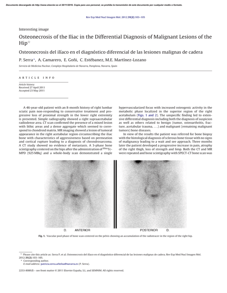
Documento descargado de http://www.elsevier.es el 20/11/2016. Copia para uso personal, se prohíbe la transmisión de este documento por cualquier medio o formato. Rev Esp Med Nucl Imagen Mol. 2012;31(2):103–105 Interesting image Osteonecrosis of the Iliac in the Differential Diagnosis of Malignant Lesions of the Hip夽 Osteonecrosis del ilíaco en el diagnóstico diferencial de las lesiones malignas de cadera P. Serra ∗ , A. Camarero, E. Goñi, C. Estébanez, M.E. Martínez-Lozano Servicio de Medicina Nuclear, Complejo Hospitalario de Navarra, Pamplona, Navarra, Spain a r t i c l e i n f o Article history: Received 27 April 2011 Accepted 23 May 2011 A 46-year-old patient with an 8-month history of right lumbar sciatic pain non-responding to conservative treatment and progressive loss of proximal strength in the lower right extremity is presented. Simple radiography showed a right supraacetabular radiodense area. CT scan confirmed the presence of a mixed lesion with lithic areas and a dense aggregate which seemed to correspond to chondroid matrix. MR imaging showed a lesion of tumoral appearance in the right acetabular region circumscribing the iliac bone with characteristics of aggressiveness based on permeation and cortical rupture leading to a diagnosis of chrondrosarcoma. A CT study showed no evidence of metastasis. A 3-phase bone scintigraphy centered on the hips after the administration of 99m TcMPD (925 MBq) and a whole-body scan demonstrated a single hypervascularized focus with increased osteogenic activity in the metabolic phase localized in the superior region of the right acetabulum (Figs. 1 and 2). The unspecific finding led to extensive differential diagnosis including both the diagnosis of suspicion as well as others related to benign (tumor, osteoarthritis, fracture, acetabular trauma, . . .) and malignant (remaining malignant tumors) bone diseases. In view of the results the patient was referred for bone biopsy with the histological diagnosis of sclerous bone tissue with no signs of malignancy leading to a wait and see approach. Three months later the patient developed a progressive increase in pain, atrophy of the right thigh, loss of strength and limp. Both the CT and MR were repeated and bone scintigraphy with SPECT-CT bone scan was Fig. 1. Vascular pool phase of bone scan centered on the pelvis showing an accumulation of the radiotracer in the region of the right hip. 夽 Please cite this article as: Serra P, et al. Osteonecrosis del ilíaco en el diagnóstico diferencial de las lesiones malignas de cadera. Rev Esp Med Nucl Imagen Mol. 2012;31(2):103–105. ∗ Corresponding author. E-mail address: patricia.serra.arbeloa@navarra.es (P. Serra). 2253-8089/$ – see front matter © 2011 Elsevier España, S.L. and SEMNIM. All rights reserved. Documento descargado de http://www.elsevier.es el 20/11/2016. Copia para uso personal, se prohíbe la transmisión de este documento por cualquier medio o formato. 104 P. Serra et al. / Rev Esp Med Nucl Imagen Mol. 2012;31(2):103–105 Fig. 2. Anterior and posterior whole-body scan showing a single augmentation of osteogenic activity in the upper region of the right acetabulum. centered on the area of interest (Fig. 3), with the results superposed to the previous findings. The patient underwent a second bone biopsy which identified bone necrosis with no signs of malignancy. Osteonecrosis or avascular bone necrosis is an infrequent pathologic entity which may appear idiopathically or secondary to different pathologies and/or external agents. The pathological changes are characterized by cell death in the bone due to a compromise in vascularization. The magnitude of avascular necrosis is based on the grade of circulatory compromise. The femoral head is the most frequent localization while the distal femur and humeral head are other usual sites. The astragalus and the carpal scaphoid are sometimes affected, although depending on the etiology, any bone of the skeleton may be affected.1 Fig. 3. Fused images of SPECT-CT scan did not show significant modifications compared to the study performed three months previously. Documento descargado de http://www.elsevier.es el 20/11/2016. Copia para uso personal, se prohíbe la transmisión de este documento por cualquier medio o formato. P. Serra et al. / Rev Esp Med Nucl Imagen Mol. 2012;31(2):103–105 The utility of bone scintigraphy to differentiate benign from malignant bone lesions has been evaluated. Despite its low specificity, it is generally highly reliable, and it is accepted that normal uptake of the bone radiotracer in the primitive bone tumor is indicative of benignity while a marked increase in uptake in the early and late phases of bone scintigraphy is more suggestive of a malignant lesion. Nonetheless, in the clinical practice there is a superposition in the scintigraphic appearance between benign and malignant lesions and thus, the greater utility of the technique lays in ruling out distant bone involvement. Osteonecrosis may present a pattern of uptake of similar characteristics in the bone repair phase which would include it in the wide differential diagnosis, although in this 105 case, in addition to being a very infrequent pathology in the localization described, neither the clinical suspicion nor the radiological data pointed to this entity.2,3 References 1. Lafforgue P. Pathophysiology and natural history of avascular necrosis of bone. Joint Bone Spine. 2006;73:500–7. 2. Mitjavila M, Balsa MA. Gammagrafía ósea en patología tumoral. In: Castro-Beiras JM, Oliva JP, editors. Oncología Nuclear. Madrid: Meditecnica; 2006. p. 305–14. 3. Mitjavila M, Balsa MA. Gammagrafía ósea en patología ósea benigna. In: CastroBeiras JM, Oliva JP, editors. Oncología Nuclear. Madrid: Meditecnica; 2006. p. 315–24.
