Lactobacillus kefiranofaciens sp. nov. Isolated from Kefir Grains
Anuncio
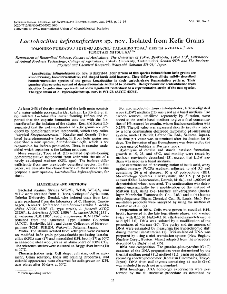
Vol. 38, No. 1 INTERNATIONAL JOURNALOF SYSTEMATIC BACTERIOLOGY, Jan. 1988, p. 12-14 0020-7713/881010012-03$02.00/0 Copyright 0 1988, International Union of Microbiological Societies Lactobacillus kefiranofaciens sp. nov. Isolated from Kefir Grains TOMOHIKO FUJISAWA,l SUSUMU ADACHL2 TAKAHIRO TOBA,* KEIZOH ARIHARA,2 AND TOMOTARI MITSUOKA1.3* Department of Biomedical Science, Faculty of Agriculture, The University of Tokyo, Bunkyo-ku, Tokyo 113'; Laboratory of Animal Products Technology, College of Agriculture, Tohoku University, Tsutsumidori, Sendai 9802;and The Institute Physical and Chemical Research, Wako-shi, Saitama 351-01, 3 Japan Lactobacillus kejiranofaciens sp. nov. is described. Four strains of this species isolated from kefir grains are slime-forming, homofermentative, rod-shaped lactic acid bacteria. They differ from all the validly described homofermentative species of the genus Lactobacilhs in their carbohydrate fermentation pattern. Their guanine-plus-cytosinecontent of deoxyribonucleicacid is 34 to 35 mol% Deoxyribonucleic acids obtained from six other Lactobacillus species do not show significant relatedness to a representative strain of the new species. The type strain of L. kejiranofaciens sp. nov. is WT-2B (ATCC 43761). . At least 24% of the dry material of the kefir grain consists of a water-soluble polysaccharide, kefiran. La Riviere et al. l(8) isolated Lactobacillus brevis forming kefiran and reported that the capsule formation was lost with the first transfer after the isolation of the strains. Rosi and Rossi (16) suggested that the polysaccharides of kefir grains are produced by homofermentative lactobacilli, which they called "atypical Streptobacterium. " Kandler and Kunath (6) isolated heterofermentative lactobacilli from kefir grains and described a new species, Lactobacillus kefir, which is not responsible for kefiran production. Thus, it remains undecided which organism is the kefiran producer. More recently, Toba et al. (17) isolated capsule-forming liomofermentative lactobacilli from kefir with the aid of a newly developed medium (KPL agar). The isolates differ sufficiently from any previously described species. In this paper, we describe the characteristics of these isolates and propose a new species, Lactobacillus kefiranofaciens, for ihem. MATERIALS AND METHODS Bacterial strains. Strains WT-2B, WT-8, WTBA, and WT-7 were obtained from T. Toba, College of Agriculture, Tohoku University, Sendai, who isolated them from kefir grain purchased from the laboratory of C. Hansen, Copenhagen, Denmark. Reference Lactobacillus strains L. acidophilus ATCC 435ST (T, type strain), L . jensenii ATCC ;!525ST, L . helveticus ATCC 15009T,L. gasseri JCM 1131T, L. crispatus JCM 1185T, and L . amylovorus JCM 1126Twere obtained from the American Type Culture Collection (ATCC), Rockville, Md., and Japan Collection of Microorganisms (JCM), RIKEN, Wako-shi, Saitama, Japan. Media. The strains isolated from kefir grain were cultured on modified kefir grain polysaccharide-producing lactobad l u s (KPL) agar (17) and in modified KPL broth (17) at 30°C in anaerobic steel wool jars in an atmosphere of 100% CO,. The reference strains were cultured on Briggs liver broth (13) at 37°C. Characterization tests. Cell shape, cell size, cell arrangement, Gram reaction, India ink staining properties, and colonial appearance were observed for cells grown on KPL agar plates after 10 days at 30°C. * Correspondingauthor. For acid production from carbohydrates, lactose-digested whey (LDW) medium (17) was used as a basal medium. The carbon sources, sterilized separately by filtration, were added to the sterile basal medium to give a final concentration of 1%,except for esculin, whose final concentration was 0.25%. The pH value was measured directly in culture tubes by a long combination electrode (automatic pH-measuring system, model BIS-120; Lifetec Co. Ltd., Saitama, Japan). The final pH value was determined after incubation for 10 days. The formation of gas from glucose was detected by the appearance of bubbles in Durham tubes. Hydrolysis of esculin and starch, catalase formation, growth at 15, 25, and 45"C, and motility were tested by methods previously described (13), except that LDW medium was used as a basal medium. For determination of the configuration of lactic acid, whey optical rototary (WOR) medium, adjusted to pH 5.5 and containing 20 g of glucose, 10 g of polypeptone (BBL Microbiology Systems, Cockeysville, Md.) 5 g of yeast extract (Difco Laboratories, Detroit, Mich.), and 1,000 ml of deproteinized whey, was used. The configuration was determined enzymatically by a modification of the method of Mattson (12), using D-( -)-lactate dehydrogenase (Boehringer Mannheim Yamanouchi Co., Ltd.) and L-( +)-lactate dehydrogenase (Sigma Chemical Co., St. Louis, Mo.). Fermentation products were analyzed by using the method of Holdeman et al. (4). Preparation of DNA. Cells were grown in modified KPL broth, harvested in the late logarithmic phase, and washed twice with 0.15 M NaCl-O.l M ethylenediaminetetraacetic acid (pH 8.0). DNA was isolated by a modification of the procedures of Marmur (10). The purity and the amount of DNA were estimated by measuring the hyperchromic shift during thermal denaturation (1). Tritium-labeled DNA was prepared by using a nick translation system (New England Nuclear Corp., Boston, Mass.) adapted from the procedure described by Rigby et al. (15). DNA base composition. The guanine-plus-cytosine (G + C) contents of the DNA preparations were determined by the thermal melting point (T,) method (ll),using an automatic recording spectrophotometer (Komatsu Electronics, Tokyo, Japan). DNA from calf thymus containing 42 mol% G+C was included in each set as a standard. DNA homology. DNA homology experiments were performed by the S1 nuclease procedure as described by Downloaded from www.microbiologyresearch.org by IP: 78.47.27.170 On: Mon, 21 Nov 2016 00:18:58 LACTOBACILLUS KEFIRANOFACIENS SP. NOV. VOL. 38, 1988 13 FIG. 1. L . kefiranofaciens sp. nov. WT-2BT, the type strain. Cells from a 10-day-old surface colony on modified KPL agar culture. Bars, 10 km. (a) Gram stain; (b) India ink stain. Johnson et al. ( 5 ) . S1 nuclease digestions were conducted with 0.5 U of S1 nuclease (Tokyo Kasei Ltd., Tokyo, Japan). After incubation for 15 min at 37"C, the remaining doublestranded DNA segments were precipitated with cold 10% trichloroacetic acid and collected on a membrane filter (type HA; Millipore Corp., Bedford, Mass.). The membranes were dried, and the radioactivity was measured in toluenebased scintillation fluid with a liquid scintillation counter (model 3330; Packard Instrument Co., Rockville, Md.). RESULTS AND DISCUSSION Morphology of the isolates. The cellular appearance of strains of the new species L. kejiranofaciens is shown in Fig. 1. All isolates grew on KPL agar at 30"C, but not on BL agar (14) and MRS agar (9). The cells were gram-positive, nonmotile, and nonsporeforming long rods (0.8 to 1.2 pm in width and 3.0 to 20.0 pm in length) and occurred singly, in pairs, or occasionally in short chains. India ink preparations of the isolates showed capsules surrounding the rods. After incubation on modified KPL agar plates (pH 5 . 5 ) for 10 days, colonies of all isolates were 0.5 to 3.0 mm in diameter, convex, transparent to translucent, entire, smooth to rough, ropy, and white (Fig. 2). Biochemical and physiological characteristics. All isolates fermented glucose, fructose, galactose, sucrose, maltose, lactose, melibiose, and raffinose. Arabinose, xylose, rhamnose, ribose, cellobiose, trehalose, melezitose, dextrin, mannitol, sorbitol, esculin, salicin, and amygdalin were not fermented. Strain WT-2B failed to ferment mannose. The final pH in glucose broth was 3.5 to 3.9. The isolates produced m-lactic acid, with a marked excess of D-(-)lactic acid. There was no growth observed at 15 and 45°C. All strains were catalase negative and did not produce gas from glucose. DNA base composition and homology. Table 1 lists the G+C contents and DNA homologies of two strains of L . kejiranofaciens examined by the S1 nuclease method. Strains WT-8 and WT-2BT had a high level of DNA homology with each other. The DNAs of six other Lactobacillus species had little or no homology with strain WT-2BT. The G+C content of the DNAs from the isolates is 34.3 to 35.4 mol%. A comparison of the biochemical and physiological characteristics, morphology, DNA base composition, and DNA homology of these isolates with those of the previously described species of the genus Lactobacillus indicate that these strains represent a new species, for which we propose the name Lactobacillus kejiranofaciens sp. nov. The phenotypic characteristics differentiating L . kejiranofaciens from other homofermentative Lactobacillus species having low G+C contents and not growing at 15°C are shown in Table 2. FIG. 2. Ten-day-old surface colonies of L . kefiranofaciens WT2BT on modified KPL agar. Bar, 5 mm. Although the habitat of L. kejir Kandler and Kunath (6) is also kefir grains, L . kejir is heterofermentative and has a G+C content of 41 to 42 mol%. Thus, L . kejir is unlike L . kejiranofaciens, which is homofermentative and has a significantly different G+C content of 34.3 to 35.4 mol%. Description of Lactobacillus kejiranofaciens sp. nov. Lactobacillus kejiranofaciens (ke.fi.rano. fa'ci.ens. L. n. kejiran, a polysaccharide of kefir grain, kefiran; L. v. facio, produce; M. L. part. adj . kejiranofaciens, kefiran producing). Gram-positive, nonmotile, capsulated, nonsporeforming rods (generally 0.8 to 1.2 pm by 3.0 to 20.0 pm) that occur singly, in pairs, or occasionally in short chains. Grows on modified KPL agar (pH 5.5) at 30°C in 10 days, producing colonies that are circular or irregular, 0.5 to 3.0 mm in diameter, convex, transparent to translucent, white, smooth to rough, and ropy. There is no growth at 15 or 45°C. Facultatively anaerobic and produces DL-lactic acid, with a marked excess of D-( -)-lactic acid homofermentatively. Catalase is not produced. Esculin and starch are not hydrolyzed. Milk is curdled. Gas is not produced from glucose. Acid is produced from glucose, fructose, galactose, sucrose, maltose, lactose, melibiose, and raffinose but not from arabinose, xylose, rhamnose, ribose, cellobiose, trehalose, melezitose, dextrin, mannitol, sorbitol, esculin, salicin, or amygdalin. DNA G+C content is 34.3 to 35.4 mol%. The habitat of the species is kefir grains. The type strain is strain WT-2BT (ATCC 43761). . TABLE 1. DNA homologies among the strains of L . kefiranofaciens and some other Lactobacillus species having low G+C contents Unlabeled DNA from strain: L . kefiranofaciens WT-2BT WT-8 L . acidophilus ATCC 4356T L. gasseri JCM 1131T L. crispatus JCM 1185T L. amylovorus JCM 1126= L . helveticus ATCC 15009= L . jensenii ATCC 25258= ~~~ ~ ~ NT, Not tested. Downloaded from www.microbiologyresearch.org by IP: 78.47.27.170 On: Mon, 21 Nov 2016 00:18:58 % Homology with reference DNA from strain WT-2BT 100 87 9 3 13 11 17 2 G + 34.3 35.4 32.3 32.9 35.4 NT" NT NT ~ ~ ~ 14 FUJISAWA ET AL. INT. J. SYST.BACTERIOL. TABLE 2. Phenotypic characteristic differentiating L . kefiranofaciens from other homofermentative Lactobacillus species that have low G+C contents and that do not grow at 15°C Reactiona L . kefiranofaciens L . acidophilus' L . amylovorus" L . crispatusc .L. gasseri" L . helveticus" .L. jensenii' .L.salivarius' L . vitulinus' L . aviariusd L . acetotoleranse a 34-35 34-37 4041 35-38 33-35 3840 35-37 34-36 3437 39-43 35-37 + + + + + + + - + - + d + + + + + - + + + d d + + + + - - d - d d + + d + + + + d - - d - d - d + - d - d d d - + + + + - - + - - - d - + + W + + - + d w - + - + W + + + d + d + - + + + + + + + + d - - + W + + + + - + -t,90% or more of strains positive; -, 90% or more strains negative; d, 11 to 89% of strains positive; w, positive to weakly positive. Based on the content of L-(+)-lactic acid as follows: D, 0 to 20%; D(L),20 to 40%; DL, 40 to 60%; L, 80 to 100%. Data from reference 7. Data from reference 3. Data from reference 2. LITERATURE CITED 1. Doty, P., J. Marmur, G. Eiger, and C. Shildraut. 1960. Strand separation in deoxyribonucleic acid: physical chemical studies. Proc. Natl. Acad. Sci. USA 46:461476. 2. Entani, E., H. Masai, and K. Suzuki. 1986. Lactobacillus acetotolerans, a new species from fermented vinegar broth. Int. J. Syst. Bacteriol. 36544549. 3. Fujisawa, T., S. Shirasaka, J. Watabe, and T. Mitsuoka. 1984. Lactobacillus aviarius sp. nov.: a new species isolated from the intestine of chickens. Syst. Appl. Microbiol. 5414420. 4. Holdeman, L. V., E. P. Cato, and W. E. C. Moore (ed.). 1977. Anaerobic laboratory manual, 4th ed. Virginia Polytechnic Institute and State University, Blacksburg. 5. Johnson, J. L., C. F. Phelps, C. S. Cummins, J. London, and F. Gasser. 1980. Taxonomy of the Lactobacillus acidophilus group. Int. J. Syst. Bacteriol. 3053-68. 6. Kandler, O., and P. Kunath. 1983. Lactobacillus kefir a component of the microflora of kefir. Syst. Appl. Microbiol. 4:28&294. 7. Kandler, O., and N. Weiss. 1986. Genus Lactobacillus Beijerinck 1901, 212AL, p. 1209-1234. In P. H. A. Sneath, N. S. Maier, M. E. Sharpe, and J. G. Holt (ed.), Bergey's manual of systematic bacteriology, vol. 2. The Williams & Wilkins Co., Baltimore. 8. La Rividre, J. W., P. Kooiman, and K. Schmidt. 1967. Kefiran, a novel polysaccharide produced in the kefir grain by Lactobacillus brevis. Arch. Microbiol. 59:269-278. 9. Man, J. C. de., M. Rogosa, and M. E. Sharpe. 1960. A medium for the cultivation of lactobacilli. J. Appl. Bacteriol. 23:130135. 10. Marmur, J. 1961. A procedure for the isolation of deoxyribonucleic acid from microorganisms J . Mol. Biol. 3:20&218. 11. Marmur, J., and P. Doty. 1962. Determination of the base composition of deoxyribonucleic acid from its thermal denaturation temperature. J. Mol. Biol. 5109-118. 12. Mattson, S. 1965. The determination of the configuration of lactic acid in bacterial cultures. Report no. 71. Milk and Dairy Research, Alnarp, Sweden. 13. Mitsuoka, T. 1969. Vergleichende Untersuchungen iiber die Laktobazillen aus den Faeces von Menschen, Schweinen und Hiihnern. Zentralbl. Bakteriol. Parsitenkd. Infektionskr. Hyg. Abt. 1 Orig. 210:32-51. 14. Mitsuoka, T., T. Sega, and S. Yamamoto. 1964. Eine verbesserte Methodik der qualitativen und quantativen Analyse der Darmflora von Menschen und Tieren. Zentralbl. Bakteriol. Parasitenkd. Infektionskr. Hyg. Abt. 1 Orig. Reihe A 195445469. 15. Rigby, P. W., M. Dieckmann, C. Rhode, and P. Berg. 1977. Labeling deoxyribonucleic acid to high specific activity in vitro by nick translation with DNA polymerase 1. J. Mol. Biol. 113: 237-251. 16. Rosi, J., and J. Rossi. 1978. I microorganismi del kefir: i fermenti lattici. Sci. Tech. Lattiero-Casearia 29:291-305. 17. Toba, T., S. Abe, K. Arihara, and S. Adachi. 1986. A medium for the isolation of capsular bacteria from kefir grains. Agric. Biol. Chem. 50:2673-2674. Downloaded from www.microbiologyresearch.org by IP: 78.47.27.170 On: Mon, 21 Nov 2016 00:18:58
