entered in the space during systole, and its exit in diastole
Anuncio
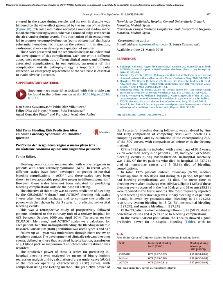
Documento descargado de http://www.elsevier.es el 21/11/2016. Copia para uso personal, se prohíbe la transmisión de este documento por cualquier medio o formato. Scientific letters / Rev Esp Cardiol. 2016;69(5):520–530 entered in the space during systole, and its exit in diastole was hindered by the valve effect generated by the suction of the device (Figure 2B). External examination revealed a rounded shadow in the blood chamber during systole, whereas a rounded bulge was seen in the air chamber during systole. This mechanism of air entrapment led to progressive pump dysfunction (pump obstruction) that had a substantial hemodynamic impact on the patient. In this situation, cardiogenic shock can develop in a question of minutes. The 2 cases presented and the schematics help us to understand the development of this complication, each one with a different appearance on examination, different clinical course, and different associated complications. In our opinion, awareness of this complication and its pathophysiology is important for early detection, as emergency replacement of the ventricle is essential to avoid adverse outcomes. SUPPLEMENTARY MATERIAL Supplementary material associated with this article can be found in the online version at doi:10.1016/j.rec.2016. 01.021. Iago Sousa Casasnovas,a,* Pablo Dı́ez Villanueva,a Felipe Dı́ez del Hoyo,a Manuel Ruiz Fernández,b Ángel González Pinto,b and Francisco Fernández-Avilésa Mid Term Bleeding Risk Prediction After an Acute Coronary Syndrome: An Unsolved Question Predicción del riesgo hemorrágico a medio plazo tras un sı´ndrome coronario agudo: una asignatura pendiente To the Editor, Bleeding complications are associated with worse prognosis in patients with acute coronary syndrome (ACS).1 In recent years, different scales have been developed to predict in-hospital bleeding complications in ACS,2–4 and these scales have been shown to have acceptable predictive power in different scenarios.5 However, these scales have not been validated for predicting bleeding complications outside the hospital setting. The objective of this study was to assess prediction of bleeding by the CRUSADE,2 Mehran,3 and ACTION4 bleeding risk scales 1 year after hospital discharge and to compare the predictive power with that shown by the 3 scales for predicting in-hospital bleeding events. This was a retrospective study of prospectively followed patients admitted to the coronary unit of a tertiary hospital for ACS between October 2009 and April 2014. The scores on the CRUSADE,2 Mehranm,3 and ACTION4 scales were calculated for each patient. To define in-hospital bleeding, the Bleeding Academic Research Consortium (BARC) definition was used (types 3 and 5).6 Follow-up at 1 year was undertaken through chart review or telephone contact. The development of clinically relevant bleeding events, defined as those that required hospitalization, transfusion of 1 blood pack, or suspension of antithrombotic treatment, was recorded. The predictive power of these 3 scales for predicting inhospital bleeding was analyzed by means of binary logistic regression analysis and by calculation of areas under curve (AUCs) of the receiver operating characteristics (ROC) curves, with comparison using the DeLong method. The predictive power of 527 a Servicio de Cardiologı´a, Hospital General Universitario Gregorio Marañón, Madrid, Spain b Servicio de Cirugı´a Cardiaca, Hospital General Universitario Gregorio Marañón, Madrid, Spain * Corresponding author: E-mail address: iagosousa@yahoo.es (I. Sousa Casasnovas). Available online 21 March 2016 REFERENCES 1. Kirklin JK, Naftel DC, Pagani FD, Kormos RL, Stevenson LW, Blume ED, et al. Sixth INTERMACS annual report: a 10,000-patient database. J Heart Lung Transplant. 2014;33:555–64. 2. Schmid C, Tjan T, Etz C, Welp H, Rukosujew A, Klotz S, et al. The Excor device, revival of an old system with excellent results. Thorac Cardiovasc Surg. 2006;54:393–9. 3. Slaughter MS, Rogers JG, Milano CA, Russell SD, Conte JV, Feldman D, et al. Advanced heart failure treated with continuous-flow left ventricular assist device. N Engl J Med. 2009;361:2241–51. 4. Hernández-Pérez FJ, Burgos-Lázaro RJ, Gómez-Bueno MF. Una complicación grave en paciente con asistencia ventricular. Rev Esp Cardiol. 2014;67:322. 5. Volz S, Holmberg M, Redfors B, Dellgren G. Acute tamponade of the left paracorporeal pump house due to membrane defect in a patient with a Berlin Heart EXCOR biventricular assist device. Eur J Cardiothorac Surg. 2014;46:743–4. 6. Pektok E, Kucukaksu S. Pulsatile paracorporeal pump membrane rupture: clinical advantage of a multilayer membrane. Artif Organs. 2012;36:936. http://dx.doi.org/10.1016/j.rec.2016.01.021 the 3 scales for bleeding during follow-up was analyzed by Fine and Gray comparison of competing risks (with death as a competing event), and by calculation of the corresponding AUC of the ROC curves, with comparison as before with the DeLong method. Of the 1489 patients included, with a mean age of 62.5 years, 77.7% were men. Forty-nine patients (3.3%) had type 3 or 5 BARC bleeding events during hospitalization. In-hospital mortality was 6.3%. Of the 94 patients who died in hospital, 35 (37.2%) died of noncardiac causes and 5 (5.3%) died of bleeding complications. In total, 1375 patients entered follow-up (97.9%, median follow-up time of 365 days), and during this period, 69 patients had bleeding complications and 73 died. The mean time to bleeding event after discharge was 169 days. Eight (11.6%) of these bleeding events occurred in the first 30 days, and 38 events (55.1%) were reported in the first 6 months. The most frequently reported type of bleeding after discharge was urinary bleeding in 24 patients (34.8%), followed by gastrointestinal bleeding in 16 (23.2%), respiratory system bleeding in 15 (21.7%), intracranial bleeding in 5 (7.2%), and muscle bleeding in 5 (7.2%). Of the 73 patients who died during follow-up, 43 (58.9%) died of noncardiac causes and 4 (5.5%) due to bleeding complications. In the overall patient population, the 3 scales showed a good predictive power for in-hospital bleeding (Table), with no Table Area Under Curve of Different Scales for Predicting Bleeding Events Scale In-hospital bleeding (AUC [95%CI]) Bleeding during follow-up (AUC [95%CI]) CRUSADE 0.75 (0.67-0.82) 0.58 (0.51-0.65) Mehran 0.71 (0.65-0.78) 0.58 (0.50-0.65) ACTION 0.73 (0.65-0.81) 0.57 (0.50-0.64) AUC, area under ROC curve; CI, confidence interval. Documento descargado de http://www.elsevier.es el 21/11/2016. Copia para uso personal, se prohíbe la transmisión de este documento por cualquier medio o formato. Scientific letters / Rev Esp Cardiol. 2016;69(5):520–530 1.00 0.50 1.00 Sensitivity A Sensitivity 528 0.00 0.50 0.00 0.00 0.50 1.00 0.00 1.00 0.50 0.50 0.00 0.00 0.50 1.00 0.00 1.00 1.00 Sensitivity Sensitivity 0.50 0.50 1 - specificity 1 - specificity 1.00 1.00 1.00 0.00 C 0.50 1 - specificity Sensitivity B Sensitivity 1 - specificity 0.00 0.50 0.00 0.00 0.50 1.00 1 - specificity 0.00 0.50 1.00 1 - specificity Figure. Receiver operating characteristics (ROC) curves for predicting in-hospital bleeding (left) and during follow-up (right). A: CRUSADE; B: ACTION; C: Mehran. statistically significant differences between the different AUCs (P not significant). By contrast, the predictive power of the 3 scales for bleeding complications at 1 year after discharge in those who survived their stay in hospital was deficient, and as before there were no significant differences between the 3 AUCs (P not significant). The Figure shows the ROC curves for predicting in-hospital bleeding events during follow-up for the 3 scales. Our study is subject to the limitations inherent in a singlecenter register, with a relatively low number of events and a homogeneous clinical management. Thus, our findings may not be applicable to populations with different characteristics and treatments. In addition, the fact that these patients are admitted to the coronary unit might imply a selection bias. The use of different bleeding definitions for the in-hospital phase and the follow-up phase after discharge is another limitation of the study. Nevertheless, in our opinion, the use of different definitions is justified by the conceptual and pathophysiological differences between in-hospital bleeding. For example, some characteristics of the definition used for bleeding after discharge (essentially, the need for hospitalization) are not applicable to a patient already in hospital. In any case, the use of different definitions cannot, we believe, explain the marked differences in AUC apparent across all 3 scales. Recent data suggest that major bleeding after discharge for ACS is associated with mortality to the same extent as during hospitalization.1 The prediction of bleeding after discharge is extremely relevant for several other reasons, such as the availability of new and potent antiplatelet drugs, doubts as to the optimal duration of dual antiplatelet therapy after ACS, or the increasingly frequent indication of anticoagulation for atrial fibrillation or other diseases due to the progressive aging of the population. In the absence of other tools, these risk scales are the ones usually used by clinicians to select the type and duration of antithrombotic treatment after discharge. Even considering the aforementioned limitations, our data show the relatively poor performance of these scales for predicting the development of bleeding complications in the first year after ACS. In our opinion, this highlights the need for new more precise and reliable tools to stratify bleeding risk after hospitalization. Documento descargado de http://www.elsevier.es el 21/11/2016. Copia para uso personal, se prohíbe la transmisión de este documento por cualquier medio o formato. Scientific letters / Rev Esp Cardiol. 2016;69(5):520–530 CONFLICTS OF INTEREST E. Abu-Assi is Associate Editor of Revista Española de Cardiologı´a. 2. Alberto Garay,a Albert Ariza-Solé,a,* Emad Abu-Assi,b Victòria Lorente,a José C. Sánchez-Salado,a and Ángel Cequiera 3. a Servicio de Cardiologı´a, Hospital Universitario de Bellvitge, L’Hospitalet de Llobregat, Barcelona, Spain b Servicio de Cardiologı´a, Complejo Hospitalario Universitario de Santiago de Compostela, Santiago de Compostela, A Coruña, Spain 4. 5. * Corresponding author: E-mail address: aariza@bellvitgehospital.cat (A. Ariza-Solé). Available online 22 March 2016 REFERENCES 1. Kazi DS, Leong TK, Chang TI, Solomon MD, Hlatky MA, Go AS. Association of spontaneous bleeding and myocardial infarction with long-term mortality Symptomatic Bradycardia and Heart Failure Triggered by Ivabradine in a Patient Receiving Antiretroviral Therapy Bradicardia sintomática e insuficiencia cardiaca precipitadas por ivabradina a una paciente que recibe tratamiento antirretroviral To the Editor, Thanks to advances in highly effective antiretroviral therapy (ART), human immunodeficiency virus (HIV) infection has become a chronic disease. HIV-infected patients now have a lifeexpectancy close to that of the general population and are increasingly affected by cardiovascular diseases, mainly ischemic heart disease and heart failure.1 ART includes drugs that are unfamiliar to most physicians, and patients sometimes do not recall the details of their medication; moreover, cardiologists are not always throroughly familiar with their patients’ ART or its possible interactions with drugs used routinely in cardiology, thereby increasing safety risks. We present the case of a 50-year-old woman with a history of essential hypertension and acute myocardial infarction, chronic heart disease with a residual left ventricular ejection fraction of 30%, and virologically and immunologically stable HIV infection (viral load < 20 copies/mL and 1069 CD4 lymphocytes/mL). Her ART included atazanavir, ritonavir, and tenofovir-emtricitabine. She was also taking carvedilol (12.5 mg/12 h), eplerenone (25 mg/24 h), ramipril (2.5 mg/24 h), and aspirin (100 mg/24 h). During an outpatient consultation, her cardiologist decided to add ivabrandine (5 mg/12 h) to her regular treatment in order to improve heart rate control. Within 48 h of starting this treatment, the patient developed a general malaise, intense astenia, and dyspnea at rest. She presented to the emergency room, where a physical examination revealed blood pressure of 80/60 mmHg, a heart rate of 45 bpm, general hypoventilation, and bilateral crackles in the lung bases. Analytical explorations gave normal results for a complete blood count, renal function, and sodium and potassium levels. A plain chest x-ray revealed signs of heart failure, and an electrocardiography examination detected sinus bradycardia at 45 bpm. After admission, the patient’s treatment for heart failure was reinforced 6. 529 after percutaneous coronary intervention. J Am Coll Cardiol. 2015;65: 1411–20. Subherwal S, Bach RG, Chen AY, Gage BF, Rao SV, Newby LK, et al. Baseline risk of major bleeding in non-ST-segment-elevation myocardial infarction: the CRUSADE (Can Rapid risk stratification of Unstable angina patients Suppress ADverse outcomes with Early implementation of the ACC/AHA Guidelines) bleeding score. Circulation. 2009;119:1873–82. Mehran R, Pocock SJ, Nikolsky E, Clayton T, Dangas GD, Kirtane AJ, et al. A risk score to predict bleeding in patients with acute coronary syndromes. J Am Coll Cardiol. 2010;55:2556–66. Mathews R, Peterson ED, Chen AY, Wang TY, Chin CT, Fonarow GC, et al. Inhospital major bleeding during ST-elevation and non-ST-elevation myocardial infarction care: derivation and validation of a model from the ACTION RegistryGWTG. Am J Cardiol. 2011;107:1136–43. Sánchez-Martı́nez M, López-Cuenca A, Marı́n F, Flores-Blanco PJ, Garcı́a Narbon A, de las Heras-Gómez I, et al. Red cell distribution width and additive risk prediction for major bleeding in non-ST-segment elevation acute coronary syndrome. Rev Esp Cardiol. 2014;67:830–6. Mehran R, Rao SV, Bhatt DL, Gibson CM, Caixeta A, Eikelboom J, et al. Standardized bleeding definitions for cardiovascular clinical trials: a consensus report from the bleeding academic research consortium. Circulation. 2011;123:2736–47. http://dx.doi.org/10.1016/j.rec.2016.02.003 and ivabradine was withdrawn. The symptoms disappeared and the patient was released from hospital after 72 h. None of the electrocardiogram traces showed evidence of QT interval prolongation. Patients infected with HIV often take protease inhibitors, among them ritonavir and nelfinavir, that are also potent inhibitors of cytochrome P450 3A4 (CYP3A4).2 Ivabradine is metabolized exclusively via this route, and therefore coadministration with protease inhibitors can considerably increase its plasma concentration, resulting in the development of excessive bradycardia, hypotension, and heart failure. Because of this interaction —described in the summary of product characteristics of protease inhibitors and ivabradine— combined treatment with these drugs is contraindicated. In any event, we have found no other report of a case similar to the one described here. Other drugs frequently used in cardiology that are contraindicated in conjunction with protease inhibitors are amiodarone (except with atazanavir), flecainide, propafenone, rivaroxaban, lovastatin, simvastatin, and lercanidipine.3,4 Coadministration with apixaban and ticagrelor should also be avoided because interactions with protease inhibitors can increase the serum concentrations of these drugs.5 Cardiologists who treat patients infected with HIV should have a thorough knowledge of their patients’ ART regimens and the possible interactions with the drugs they commonly prescribe. On its website, GESIDA (Grupo de Estudio del SIDA de la Sociedad Española de Enfermedades Infecciosas y Microbiologı´a Clı´nica [AIDS Study Group, Spanish Society of Infectious Diseases and Clinical Microbiology]) provides numerous very useful action guidelines; of particular relevance to cardiologists is the ‘‘Documento de consenso sobre alteraciones metabólicas y riesgo cardiovascular en pacientes con infección por el VIH (Febrero 2014) (Consensus document on metabolic alterations and cardiovascular risk in patients infected with HIV [February 2014])’’.6 José M. Romero-León,* Marı́a C. Gálvez-Contreras, and Luis F. Dı́ez-Garcı́a Unidad de Enfermedades Infecciosas, Servicio de Medicina Interna, Complejo Hospitalario Torrecárdenas, Almerı´a, Spain
