Invasion of the Spinal Column by a Posterior Mediastinal Cavernous
Anuncio
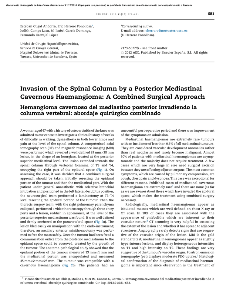
Documento descargado de http://www.elsevier.es el 21/11/2016. Copia para uso personal, se prohíbe la transmisión de este documento por cualquier medio o formato. cir esp. 2013;91(10):677–691 Esteban Cugat Andorra, Eric Herrero Fonollosa*, Judith Camps Lasa, M. Isabel Garcı́a Domingo, Fernando Carvajal López *Corresponding author. E-mail address: eherrero@mutuaterrassa.es (E. Herrero Fonollosa). Unidad de Cirugı́a Hepatobiliopancreática, Servicio de Cirugı́a General, Hospital Universitari Mutua de Terrassa, Tarrasa, Universitat de Barcelona, Spain 2173-5077/$ – see front matter # 2012 AEC. Published by Elsevier España, S.L. All rights reserved. 681 Invasion of the Spinal Column by a Posterior Mediastinal Cavernous Haemangioma: A Combined Surgical Approach Hemangioma cavernoso del mediastino posterior invadiendo la columna vertebral: abordaje quirúrgico combinado A woman aged 67 with a history of osteoarthritis of the knee was admitted to our centre to investigate a clinical history of weeks of difficulty in walking, dysaesthesia in both lower limbs and pain at the level of the spinal column. A computerised axial tomography scan (CT) and magnetic resonance imaging (MRI) were performed which revealed a well-defined 39 mm38 mm lesion, in the shape of an hourglass, located at the posterior superior mediastinal level. The lesion extended towards the spinal column through vertebral foramina of T3 and T4, occupying the right part of the epidural space (Fig. 1). On assessing the case, it was decided that a combined surgical approach should be taken, initially resecting the epidural portion of the tumour and then the mediastinal part. With the patient under general anaesthetic, with selective bronchial intubation and positioned in the left lateral decubitus position, the neurosurgical team performed a laminectomy at T3–T4 level resecting the epidural portion of the tumour. Then the thoracic surgery team, with the right pulmonary parenchyma collapsed, performed a videothoracoscopy through three entry ports and a lesion, reddish in appearance, at the level of the posterior superior mediastinum was found. It was well defined and firmly anchored to the paravertebral space (Fig. 2a). The lesion bled easily on manipulation with the endo-instrument; therefore, an auxiliary anterior minithoracotomy was performed to free the mass safely. Once the tumour had been freed a communication orifice from the posterior mediastinum to the epidural space could be observed, created by the growth of the tumour. The anatomo-pathological study showed that the epidural portion of the tumour measured 13 mm1 mm and the mediastinal portion was encapsulated and measured 35 mm2 mm25 mm. The tumour was compatible with a cavernous haemangioma (Fig. 2b). The patients had an § uneventful post-operative period and there was improvement of the symptoms on admission. Mediastinal haemangiomas are extremely rare tumours with an incidence of less than 0.5% of all mediastinal tumours. They are considered vascular development anomalies rather than real neoplasias and rarely become malignant. Almost 50% of patients with mediastinal haemangiomas are asymptomatic and the majority does not require treatment. A few cases which are very large in size need surgical excision because they are affecting adjacent organs. The most common symptoms, which are caused by pulmonary compression, are cough, chest pain and dyspnoea. This case was exceptional for different reasons. Published cases of mediastinal cavernous haemangioma are extremely rare1 and there are none (as far as we are aware) about those which have invaded the epidural space, which makes the treatment using combined surgery necessary. Radiologically, mediastinal haemangiomas appear as lobulated masses which are well defined on chest X-ray or CT scan. In 10% of cases they are associated with the appearance of phleboliths which are inherent to their vascular nature.2 CT scanning is very helpful in assessing the extent of the lesion and whether it has spread to adjacent structures. Angiography rarely detects signs that are suggestive of the vascular origin of the lesion. MRI is the gold standard test; mediastinal haemangiomas appear as slightly hyperintense lesions, and display heterogeneous intensities on T1 and high intensity on T2. These findings are very suggestive of the tumour’s vascular origin. Positron emission tomography (pet) displays moderate FDG uptake.3 Histological confirmation of the diagnosis of mediastinal haemangioma is important since observation is the treatment of Please cite this article as: Fibla JJ, Molins L, Mier JM, Conesa G, Garcı́a F. Hemangioma cavernoso del mediastino posterior invadiendo la columna vertebral: abordaje quirúrgico combinado. Cir Esp. 2013;91:681–683. Documento descargado de http://www.elsevier.es el 21/11/2016. Copia para uso personal, se prohíbe la transmisión de este documento por cualquier medio o formato. 682 cir esp. 2013;91(10):677–691 Fig. 1 – Magnetic resonance T2 showing a marked hyper-intensity of the mass invading the epidural space: (A) coronal section, (B) sagittal section and (C) transverse section. Fig. 2 – (A) Intra-operative findings show a well-defined, reddish mass in the posterior superior mediastinum. (B) Microscopic view showing vascular lakes with flattened endothelium. No atypias confirmed; 20T haematoxylin–eosin. Documento descargado de http://www.elsevier.es el 21/11/2016. Copia para uso personal, se prohíbe la transmisión de este documento por cualquier medio o formato. cir esp. 2013;91(10):677–691 choice for asymptomatic lesions as they might resolve spontaneously. Nonetheless, the progression of the tumour needs to be observed as there are two histological varieties of mediastinal haemangioma: capillary and cavernous. Both varieties display different growth patterns.4 Unlike capillary haemangiomas, mediastinal cavernous haemangiomas do not resolve spontaneously. The problem is that it is very difficult to make a histological diagnosis of these tumours using methods which are not invasive. The method of diagnosis and treatment of choice is complete resection of the tumour. Haemorrhage is the main risk factor in surgical excision of these tumours. In cases where radical resection is not feasible, radiotherapy has been used as an alternative treatment.5 683 3. Sakurai K, Hara M, Ozawa Y, Nakagawa M, Shibamoto Y. Thoracic hemangiomas: imaging via CT, MR, and PET along with pathologic correlation. J Thorac Imaging. 2008;23:114–20. 4. Moran CA, Suster S. Mediastinal hemangiomas: a study of 18 cases with emphasis on the spectrum of morphological features. Hum Pathol. 1995;26:416. 5. Cohen AJ, Sbaschnig RJ, Hochholzer L, Lough FC, Albus RA. Mediastinal hemangioma. Ann Thorac Surg. 1987;43: 656–9. Juan J. Fiblaa,*, Laureano Molinsa, José M. Miera, Gerardo Conesab, Felip Garcı́ac a Servicio de Cirugı́a Torácica, Hospital Quirón, Barcelona, Spain Servicio de Neurocirugı́a, Hospital Quirón, Barcelona, Spain c Servicio de Anatomı́a Patológica, Hospital Quirón, Barcelona, Spain b references 1. Ampollini L, Carbognani P, Cattelani L, Bilancia R, Rusca M. Cavernous hemangioma of the posterior mediastinum. Ann Thorac Surg. 2010;90:e96. 2. Hanaoka J, Inoue S, Fujino S, Kontani K, Sawai S, Tezuka N, et al. Mediastinal cavernous hemangioma in a child: report of a case. Surg Today. 2002;32:985–8. *Corresponding author. E-mail address: juanjofibla@gmail.com (J.J. Fibla). 2173-5077/$ – see front matter # 2012 AEC. Published by Elsevier España, S.L. All rights reserved. ‘‘Second-Look’’ in Initially Unresectable Pancreatic Adenocarcinoma After Neoadjuvant Chemotherapy «Second-look» en adenocarcinoma de páncreas inicialmente irresecable tras quimioterapia neoadyuvante Surgery is the only potentially ‘‘curative’’ therapy for pancreatic cancer, which is still the most lethal gastrointestinal neoplasia.1,2 Unfortunately due to its rapid and insidious growth for years it has been highly unresectable.1 It is estimated, therefore, that around 80%–90% of cases are inoperable at the time of diagnosis, because they are locally advanced and/or metastatic, and they have an overall survival (five years) of 5%, rising to 25% in those susceptible to oncological resection.3 Although there is consensus that curative surgery is not feasible in the case of infiltration of the coeliac trunk or the superior mesenteric artery, there is less agreement about other forms of locally advanced disease. In fact, over the past few decades, the concept of resectability has evolved and there is a tendency to take a more ambitious and individualised approach in tackling this typically aggressive tumour biology. § Therefore, benefits in selected patients have now been documented with respect to neoadjuvant chemo-radiotherapy.4 In this regard, comparable overall and disease-free survival has been noted between initially and secondarily resected neoplasias as long as free margins are achieved. Indeed, in some cases, paradoxically, it has been higher in the second group (20 compared to 33 months average survival).1 However, the therapeutic strategy adopted by different groups with experience in second-look pancreatic cancer surgery varies considerably; from the most conservative policies (MSKCC) in which re-exploration is only considered when there are at least partial responses with a priori curative possibility (R0), to the policy of the University of Duke where they indicate re-laparotomy as long as there is no portal thrombosis or arterial involvement, and to the policy promoted by Massucco et al. who consider resection not only in the case of partial regressions, but also in the case of stable Please cite this article as: Gómez Garcı́a ME, Carbonell Castelló F, Alberola Soler A, Poves Gil PM, Garcı́a Espinosa R. «Second-look» en adenocarcinoma de páncreas inicialmente irresecable tras quimioterapia neoadyuvante. Cir Esp. 2013;91:683–685.
