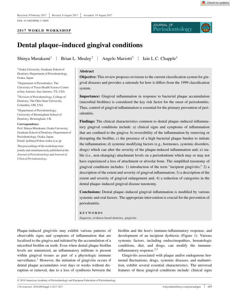
Received: 8 February 2017 Revised: 8 August 2017 Accepted: 19 August 2017 DOI: 10.1002/JPER.17-0095 2017 WORLD WORK SHOP Dental plaque–induced gingival conditions Shinya Murakami1 Brian L. Mealey2 1 Osaka University, Graduate School of Dentistry-Department of Periodontology, Osaka, Japan 2 Department of Periodontics, The Angelo Mariotti3 Iain L.C. Chapple4 Abstract Objective: This review proposes revisions to the current classification system for gingival diseases and provides a rationale for how it differs from the 1999 classification University of Texas Health Science Center at San Antonio, San Antonio, TX, USA system. 3 Division of Periodontology, College of Importance: Gingival inflammation in response to bacterial plaque accumulation Dentistry, The Ohio State University, Columbus, OH, USA (microbial biofilms) is considered the key risk factor for the onset of periodontitis. Thus, control of gingival inflammation is essential for the primary prevention of peri- 4 Department of Periodontology, University of Birmingham School of Dentistry, Birmingham, UK Correspondence Prof. Shinya Murakami, Osaka University, Graduate School of Dentistry-Department of Periodontology, Osaka, Japan. Email: ipshinya@dent.osaka-u.ac.jp The proceedings of the workshop were jointly and simultaneously published in the Journal of Periodontology and Journal of Clinical Periodontology. odontitis. Findings: The clinical characteristics common to dental plaque–induced inflammatory gingival conditions include: a) clinical signs and symptoms of inflammation that are confined to the gingiva: b) reversibility of the inflammation by removing or disrupting the biofilm; c) the presence of a high bacterial plaque burden to initiate the inflammation; d) systemic modifying factors (e.g., hormones, systemic disorders, drugs) which can alter the severity of the plaque-induced inflammation and; e) stable (i.e., non-changing) attachment levels on a periodontium which may or may not have experienced a loss of attachment or alveolar bone. The simplified taxonomy of gingival conditions includes: 1) introduction of the term “incipient gingivitis;” 2) a description of the extent and severity of gingival inflammation; 3) a description of the extent and severity of gingival enlargement and; 4) a reduction of categories in the dental plaque–induced gingival disease taxonomy. Conclusions: Dental plaque–induced gingival inflammation is modified by various systemic and oral factors. The appropriate intervention is crucial for the prevention of periodontitis. KEYWORDS diagnosis, evidence-based dentistry, gingivitis Plaque-induced gingivitis may exhibit various patterns of observable signs and symptoms of inflammation that are localized to the gingiva and initiated by the accumulation of a microbial biofilm on teeth. Even when dental plaque biofilm levels are minimized, an inflammatory infiltrate is present within gingival tissues as part of a physiologic immune surveillance.1 However, the initiation of gingivitis occurs if dental plaque accumulates over days or weeks without disruption or removal, due to a loss of symbiosis between the biofilm and the host's immune-inflammatory response, and development of an incipient dysbiosis (Figure 1). Various systemic factors, including endocrinopathies, hematologic conditions, diet, and drugs, can modify the immuneinflammatory response.2,3 Gingivitis associated with plaque and/or endogenous hormonal fluctuations, drugs, systemic diseases, and malnutrition, exhibit several essential characteristics. The universal features of these gingival conditions include: clinical signs © 2018 American Academy of Periodontology and European Federation of Periodontology J Periodontol. 2018;89(Suppl 1):S17–S27. wileyonlinelibrary.com/journal/jper S17 MURAKAMI ET AL. S18 Behavioural risk factors absent Behavioural risk factors present Environmental risk factors absent Environmental risk factors evident Clinical Health Complement Health Promong PMNs biofilm = Symbiosis Periodons Gingivis Anbody Anbody Proporonate Host response Incipient Proporonate Dysbiosis PMNs ++ Host response (Quorum Sensing Bacteria) Frank Dysbiosis (Pathogenic Biofilm) T & B cells Disproporonate PMNs +++ Host response (hyperPlasma cells inflammatory) Angens Angens Low biomass fMLP Acute Resoluon of inflammaon Virulence Factors High biomass LPS Haem ↑ GCF Connecve Tissue & Bone Damage Cytokines Angens Bact’l DNA DAMPs Chronic Resoluon of inflammaon High biomass Gingipains LPS Prostanoids Failed MMPs Chronic nonResolving Resoluon of inflammaon Oxidave inflammaon Stress Genec risk factors absent Epigenec effects not evident Genec risk factors present Chapple 2015 Epigenec effects evident FIGURE 1 Contemporary model of host–microbe interactions in the pathogenesis of periodontitis, in which the host response drives an incipient dysbiosis (gingivitis). If the biofilm is not disrupted/removed, frank dysbiosis results and perpetuates a chronic nonresolving and destructive inflammation. DAMPs, damage-associated molecular patterns; fMLP, N- formylmethionyl- leucyl- phenylalanine; GCF, gingival crevicular fluid; LPS, lipopolysaccharide; MMPs, matrix metalloproteinases; PMNs, polymorphonuclear neutrophils. This figure is referred from ref. 106. and symptoms of inflammation that are confined to the free and attached gingiva and do not extend beyond the mucogingival junction; reversibility of the inflammation by disrupting/removing the biofilm; the presence of a high bacterial plaque burden to initiate and/or exacerbate the severity of the lesion (although this varies among individuals); and stable (i.e., unchanging) attachment levels on a periodontium, which may or may not have experienced a loss of attachment or alveolar bone. Gingival inflammation is regarded as a necessary prerequisite for the subsequent development of periodontitis and progressive attachment loss around teeth.4 Management of gingivitis is therefore a key primary preventive strategy for periodontitis and a secondary preventive strategy for recurrence of periodontitis.5,6 METHODS This review updates and revises the previous classification of plaque-induced gingival conditions reported in the 1999 classification system.5 A literature search was conducted using the PubMed interface with “Gingival diseases” [MeSH] or “Gingivitis” [MeSH] and other related MeSH terms, such as “Microbiota”[MeSH], “Gonadal Steroid Hormones”[MeSH], “Hyperglycemia”[MeSH], “Dental Prosthesis”[MeSH], applied to systematic and narrative reviews as well as original research articles published after 1999. Also utilized were manual search approaches to identify additional primary studies; studies relating to non-plaque-induced gingival lesions were not considered. Observations and discussion References employed in the 1999 classification system5 were reviewed, and the appropriate ones were selected for re-analysis. In addition, papers related to “gingivitis” were retrieved using Medline and were finally selected based on the discussion of the authors and supplemented by suggestions of the co-chairs of the group. P LAQUE- INDUCED G INGIVITIS Plaque-induced gingivitis is an inflammatory response of the gingival tissues resulting from bacterial plaque accumulation located at and below the gingival margin.6 It does not directly cause tooth loss; however, managing gingivitis is a primary preventive strategy for periodontitis.7 Epidemiologic data have shown plaque-induced gingivitis to be prevalent at all ages in dentate populations,8–14 and this disease is considered the most common form of periodontal disease15 (Table 1). The initial changes from health to plaque-induced gingivitis may not be detectable clinically,16 raising important debates concerning clinical thresholds for defining physiologic vs pathologic inflammation. However, as plaque-induced gingivitis progresses to more established forms of this disease, clinical signs and symptoms become obvious. Plaque-induced gingivitis begins at the gingival margin and may spread throughout the remaining gingival unit. Patients may notice symptoms that include bleeding with tooth brushing, blood in saliva, gingival swelling and redness, and halitosis in the case of established forms.17 MURAKAMI ET AL. TABLE 1 S19 Summary of epidemiologic studies on gingivitis Age, years Sample size Method Definition Prevalence Employed adults and seniors in U.S. 18-79 6,672 National survey PI 85.5% (male) 78.8% (female) including periodontitis 1972 Children in U.S. 6-11 7,109 National survey PI 38.7% 1987 Employed adults in 18-64 U.S. 15,132 National survey Bleeding on gentle sweep of the gingival margin 47% (male) 39% (female) Retired persons in 65-80+ U.S. 5,686 Ref. Author Year Population 8 U.S. Public Health Service NCHS 1965 9 U.S. Public Health Service NCHS 11 U.S. Public Health Service NIDR 53% (male) 44% (female) 12 Bhat 1991 School children in 14-17 U.S. 11,111 National survey Bleeding on gentle sweep of the gingival margin 58.8% 15 White et al. 2012 Adults in U.K. excluding Scotland 6,469 National survey BOP, calculus, pocket depth, attachment level 83% including periodontitis 16+ PI, periodontal index; BOP, bleeding on probing TABLE 2 Classification of plaque-induced gingivitis and modi- fying factors A. Associated with bacterial dental biofilm only B. Potential modifying factors of plaque-induced gingivitis 1. Systemic conditions a) Sex steroid hormones 1) Puberty 2) Menstrual cycle 3) Pregnancy 4) Oral contraceptives b) Hyperglycemia c) Leukemia d) Smoking e) Malnutrition 2. Oral factors enhancing plaque accumulation a) Prominent subgingival restoration margins b) Hyposalivation C. Drug-influenced gingival enlargements The intensity of the clinical signs and symptoms will vary among individuals18 as well as among sites within a dentition. The common clinical signs of plaque-induced gingivitis include erythema, edema, bleeding, tenderness, and enlargement.7,19 The severity of plaque-induced gingivitis can be influenced by tooth and root anatomy, restorative and endodontic considerations, and other tooth-related factors20 (Table 2). Radiographic analysis and/or probing attachment levels of individuals with plaque-induced gingivitis will generally not indicate loss of supporting structures. Histopathologic changes include the elongation of rete ridges into the gingival connective tissue, vasculitis of blood vessels adjacent to the junctional epithelium, progressive destruction of the collagen fiber network with changes in collagen types, cytopathologic alterations of resident fibroblasts, and a progressive inflammatory/immune cellular infiltrate.16 Although recent studies suggest that bacterial phylotypes associated with gingivitis are distinct from those associated with health or periodontitis,21–24 further studies are needed to clearly define the microbial community of gingivitis. In this regard, gingivitis is a non-specific dental plaque–induced inflammatory condition, a concept that remains unchanged from 1999. The molecular characteristics or the pattern of the gingival transcriptome (i.e., sum total of all mRNA expressed by genes found in the gingiva) during plaque-induced gingival inflammation have been scrutinized since the last classification workshop. Because mRNA transcripts are not always translated into proteins, it is important to understand which transcripts are expressed as proteins25 and causally related to the onset of gingival inflammation, and which are risk factors or risk predictors of gingival inflammation. Currently, several broad biologic changes in the transcription of genes from noninflamed to inflamed gingival units have been documented, and ontologic groupings and include: 1) host-bacterial interactions, including but not limited to microbial pattern recognition molecules; 2) host cell chemotaxis; 3) phagocytosis and degranulation; 4) novel cellular/molecular pathway signaling, including but not limited to cytokine signaling and cell adhesion; 6) T lymphocyte response; 7) angiogenesis; and 8) epithelial immune response.26,27 At this time, the role of the gingival transcriptome is only beginning to be understood in relation to gingival inflammation. MURAKAMI ET AL. S20 Plaque-induced gingivitis on a reduced periodontium Following active periodontal treatment and the resolution of inflammation from periodontitis, the periodontal tissue is clinically non-inflamed but with a reduced connective tissue attachment and alveolar bone height. Plaque-induced gingivitis on a reduced periodontium is characterized by the return of bacterially induced inflammation to the gingival margin on a reduced periodontium with no evidence of progressive attachment loss (i.e., no indication of active disease). The common clinical and microbial findings are the same as plaque-induced gingivitis on a full periodontium except for the presence of pre-existing attachment loss and therefore a higher risk of periodontitis, unless professional, tailored supportive care regimens are in place.28 MODIFYING FAC TOR S O F PLAQUE-INDU C E D G I NGI V I T I S Plaque-induced gingivitis exacerbated by sex steroid hormones Homeostasis within the periodontium involves complex, multifactorial endocrine relationships.29,30 Evidence has accrued to show that tissue responses within the periodontium are modulated by androgens, estrogens, and progestins at one time or another in a person's life.29,30 For endocrinotropic conditions, plaque bacteria in conjunction with elevated steroid hormone levels are necessary to produce a gingival inflammatory response. The composition of the required flora has not been fully elucidated;31 therefore, bacteriologic analysis of endocrinotropic gingival conditions is not currently useful for diagnosis.29,30 The following conditions may modify plaque-induced gingivitis but are not considered diagnoses in and of themselves (Table 2). Menstrual cycle During the menstrual cycle, significant and observable inflammatory changes in the gingiva have been documented in case reports.36,37 However, most clinical studies have shown there are only modest inflammatory changes that may be observable during ovulation.33,34,36 More specifically, gingival crevicular fluid flow has been shown to increase by at least 20% during ovulation in over 75% of women tested,38 and other studies have also shown a modest change in women with pre-existing periodontal inflammation. Although there may be a very small cohort of women who are extremely sensitive to hormonal changes in the gingiva during the menstrual cycle, most women with menstrual cycle–associated gingival inflammation will present with clinically non-detectable signs of the condition39–41 (Table 3). Pregnancy During pregnancy, the prevalence and severity of gingivitis has been reported to be elevated and frequently unrelated to the amount of plaque present.38,42–45 Both longitudinal and cross-sectional studies have found the prevalence and severity of gingival inflammation significantly higher in the pregnant vs the post-partum patient, even though plaque scores remained the same between the two groups.38,42 Furthermore, gingival probing depths are deeper,38,42,44 bleeding on probing or bleeding with toothbrushing is also increased,42,44 and gingival crevicular fluid flow is elevated38 in pregnant women. The features of pregnancy-associated gingivitis are similar to plaque-induced gingivitis, except the propensity to develop frank signs of gingival inflammation in the presence of a relatively small amount of plaque during pregnancy. Pregnancy may also be associated with the formation of pregnancy-associated pyogenic granulomas. This topic is covered by Holmstrup et al. from this workshop.46 Puberty Oral contraceptives The incidence and severity of gingivitis in adolescents are influenced by a variety of factors, including dental plaque biofilm levels, dental caries, mouth breathing, crowding of the teeth, and tooth eruption.10 However, the dramatic rise in steroid hormone levels during puberty has a transient effect on the inflammatory status of the gingiva.29,30 A number of studies have demonstrated an increase in gingival inflammation of circumpubertal age and in both genders, without a concomitant increase in plaque levels.32–35 Although puberty-associated gingivitis has many of the clinical features of plaque-induced gingivitis, it is the propensity to develop frank signs of gingival inflammation in the presence of relatively small amounts of plaque during the circumpubertal period that are key to distinguishing this condition. Oral contraceptive agents were once associated with gingival inflammation and gingival enlargements. In the early studies, the increased gingival inflammation or enlargement was reversed when oral contraceptive use was discontinued or the dosages reduced. The features of gingivitis associated with oral contraceptives in premenopausal women were similar to plaque-induced gingivitis, except the propensity to develop frank signs of gingival inflammation in the presence of relatively small amounts of plaque in women taking these hormones. Current oral contraceptive concentrations are much lower than the original doses that were reported in these early clinical studies, and it is known that current formulations of oral contraceptive do not induce the clinical changes in gingiva that were reported with high-dose contraceptives.29,30,47 MURAKAMI ET AL. TABLE 3 S21 Summary of studies on menstrual cycle-associated gingival changes Age, years Ref. Author Year Population 39 2009 Female dental 19-23 students in Turkey 40 41 Baser et al. Becerik et al. Shourie et al. 2010 2012 Premenopausal women in Turkey Premenopausal women in India 21-40 18-40 Sample size Evaluation Outcome 27 PI, GI no change between MD, OD, PgD 50 (25 gingivitis, 25 healthy subjects) BOP more in PgD than MD and OD GCF amount more in PgD than OD IL-1𝛽, TNF-𝛼 in GCF more IL-1𝛽 in PgD than MD and OD BOP more in PM than OV and ME of gingivitis subjects PI more in gingivitis than healthy subjects; same in OV, PM, ME GCF amount more in gingivitis than healthy subjects; same in OV, PM, ME IL-6, PGE2, PAI-2, t-PA in GCF more IL-6 in gingivitis than healthy subjects; same in OV, PM, ME PI 100 (25 gingivitis, 25 gingivitis treated, 50 no existing gingivitis) identical in OV, PM, ME GI OV > PM > ME in gingivitis subjects but no difference in treated and no existing gingivitis subjects GCF amount OV > PM > ME in gingivitis subjects but no difference in treated and no existing gingivitis subjects PI: periodontal index; GCF: gingival crevicular fluid; ME: menstruation; PM: premenstruation; OV: ovulation; MD: the first menstruation day; OD: estimated ovulation day; PgD: estimated progesterone secretion day; IL, interleukin; TNF-𝛼, tumor necrosis factor-alpha; PGE2, prostaglandin E2; PAI-2, plasminogen activator inhibitor-2; t-PA, tissue plasminogen activator. PLAQUE-INDU C E D G I NGI V I T I S E X AC E R BAT E D BY SYST E M I C CON D I T I O N S Hyperglycemia, hematologic malignancies, and nutrient deficiencies are a remarkably diverse collection of systemic states that can affect the gingival tissues. For specific systemic conditions, such as hyperglycemia, acute leukemias, and/or vitamin C deficiency, plaque bacteria are necessary to produce a gingival response. Hyperglycemia Gingivitis is a consistent feature found in children with poorly controlled type 1 diabetes mellitus, and the level of glycemic control may be more important in determining the severity of gingival inflammation than the quality of plaque control.48–50 In adults with diabetes mellitus it is much more difficult to detect the effects of this endocrine disease on gingival diseases, and only limited evidence is available51 since most studies have evaluated gingival inflammation in association with attachment loss.52 Leukemia Oral manifestations have been described primarily in acute leukemia and consist of cervical lymphadenopathy, petechiae, and mucosal ulcers as well as gingival inflammation and enlargement.53 Signs of inflammation in the gingiva include swollen, glazed, and spongy tissues which are red to deep purple in appearance.54 Gingival bleeding is a common sign in patients with leukemia and is the initial oral sign and/or symptom in 17.7% and 4.4% of patients with acute and chronic leukemias, respectively.53 The bleeding is due to thrombocytopenia and clotting factor deficiencies and can present in preleukemic states such as myelodysplasia as an initial sign.55 Gingival enlargement has also been reported, initially beginning at the interdental papilla followed by the marginal and attached gingiva.53,54 The enlargement is caused by infiltration of gingivae by leukemic cells.55 Although local MURAKAMI ET AL. S22 irritants can predispose to exacerbate the gingival response in leukemia, they are not prerequisites for lesions to form in the oral cavity.54 Smoking Epidemiologic studies have revealed that smoking is one of the major lifestyle-related environmental risk factors for periodontal disease.56 Both the local and systemic effects of cigarette smoke should be intrinsically considered. Inhaled cigarette smoke is absorbed from the capillary vessels via the pulmonary alveolar epithelium and enters the systemic circulation, whereas direct exposure of inhaled cigarette smoke to periodontal tissues causes vasoconstriction of the periodontal microvasculature and gingival fibrosis, which is often observed in smokers.57 Although plaque accumulation and disease progression are exacerbated in smokers, smokers have fewer clinical signs and symptoms of gingival inflammation, and therefore smoking can mask an underlying gingivitis.58,59 Malnutrition The precise role of nutrition in the initiation or progression of periodontal diseases remains to be elucidated, leading to a paucity of information available regarding the effects of almost all nutritional deficiencies on human periodontal tissues. The one nutritional deficiency that has well-documented effects on the periodontium involves depletion of plasma ascorbic acid (i.e., vitamin C). Even though scurvy is unusual in areas with an adequate food supply, certain populations on restricted diets (e.g., infants from low socioeconomic families, the institutionalized elderly, and alcoholics) are at risk of developing this condition.60 Although there is no dispute about the necessity of dietary ascorbic acid for periodontal health,61 in the absence of frank scurvy, the effect of declining ascorbic acid levels on the gingiva can be difficult to detect clinically,62 and when it is detected, it usually has characteristics that are similar to plaque-induced gingivitis. PLAQUE-INDU C E D G I NGI V I T I S E X AC E R BAT E D BY O R A L FAC TO R S The onset and progress of gingival inflammation can be modified/exacerbated by various oral (local) factors as documented below. Prominent subgingival restoration margins The subgingival convexity and margin of a restoration is very important in site-specific plaque control and is closely related to gingival health. Although higher level clinical evidence in the field is not available, the concept that restoration margins placed apical to the gingival margin are detri- mental to gingival health has been confirmed by a 26-year longitudinal study.63 Prominent subgingival restoration margins promote gingivitis by increasing the local accumulation of bacterial plaque. Thus, subgingival restoration margins need to be carefully designed in order to minimize plaque retention. Hyposalivation Xerostomia is a symptom caused by a perceived lack of saliva in the oral cavity, rather than a diagnosis per se;64,65 hence, the term “hyposalivation” is employed here as a diagnostic term. It is known that some health conditions/diseases such as Sjögren's syndrome, anxiety, and poorly controlled diabetes may cause xerostomia due to hyposalivation. Importantly, it is frequently observed as a side effect of medications such as antihistamines, decongestants, antidepressants, antihypertensive medications. Hyposalivation may cause progressive dental caries, taste disorders, halitosis, and inflammation of the oral mucosa, tongue, and gingiva.66,67 Dryness in the mouth may make plaque control difficult, and gingival inflammation may be worsened. D RU G - I N F LU E NC E D G I NGI VA L ENLARGEMENTS There are an assortment of medications that have been reported to affect the size of the gingival tissues.68 In the literature, the drugs primarily associated with gingival tissue enlargement have included the antiepileptic drugs phenytoin and sodium valproate, certain calcium channel–blocking drugs (e.g., nifedipine, verapamil, diltiazem, amlodipine, felodipine), immunoregulating drugs (e.g., ciclosporine), and high-dose oral contraceptives.69–71 For drug-influenced gingival conditions, plaque bacteria in conjunction with the drug are necessary to produce a gingival response. Nonetheless, not all individuals who take these medications will develop enlargements of the gingival tissues, suggesting a susceptibility requiring specific characteristics.72 Furthermore, some sites/patients with drug-influenced gingival enlargement present little, if any, clinically evident gingivitis at affected sites. The common clinical characteristics of drug-influenced gingival enlargements include variations in interpatient or intrapatient patterns of enlargement (i.e., genetic predisposition),69,70 a tendency to occur more often in the anterior gingiva,69,70 a higher prevalence in younger age groups,73–75 onset within 3 months of use69,74,75 that is usually first observed at the papilla,69 and, although it can be found in a periodontium with or without bone loss, it is not associated with attachment loss or tooth mortality.69,70,76 Finally, all of these drugs produce clinical lesions and MURAKAMI ET AL. histologic characteristics that are indistinguishable from one another.69,70 R E VIS IONS TO T H E 1999 D E N TA L PLAQUE– INDU C E D G I NGI VA L DISEASES CLASSIFICATION SYST E M Plaque-induced gingivitis can arise in any individual due to an increase in biofilm accumulation, and gingivitis may be exacerbated by systemic states. From the previous 1999 taxonomy of plaque-induced gingival conditions, it is believed the classification can be simplified to represent society's current perception of disease and health, which has been influenced by our expanding scientific knowledge base as well as our cultural, social, and individual value judgments. Similar to the 1999 classification system, plaque-induced gingival inflammatory conditions require the presence of dental plaque coupled with clinical signs and symptoms of gingival inflammation in an otherwise stable periodontium. The revision of the 1999 classification system for dental plaque-induced gingival diseases involved four components: 1) description of the extent and severity of the gingival inflammation, 2) description of the extent and severity of gingival enlargements, 3) a reduction in gingival disease taxonomy, and 4) discussion of whether mild localized gingivitis should be considered a disease or variant of health. To begin, the extent, or the number of gingival sites exhibiting inflammation, can be described as either localized or generalized. Similar to the manner in which extent is described for chronic periodontitis, a gingival condition would be described as localized when < 30% of the teeth are affected, and generalized would reflect when ≥30% of the teeth are affected by gingival inflammation. In addition, it is proposed to consider introducing the term “incipient gingivitis” where, by definition, only a few sites are affected by mild inflammation, expressed as mild redness and/or a delayed and broken line of bleeding rather than edema or an immediate unbroken line of bleeding on probing. Incipient gingivitis may be regarded as a condition that is part of a spectrum of “clinical health,” but may rapidly become localized gingivitis if untreated. The severity, or intensity of inflammation at a site, tooth, or the entire dentition, would be reflected by the gingival index described by Loe (1967).77 More specifically, mild gingival inflammation would be an area with a minor change in color and little change in the texture of the tissue. Moderate gingival inflammation would be an area with glazing, redness, edema, enlargement, and bleeding upon probing; severe gingival inflammation would be an area of overt redness and S23 edema with a tendency toward bleeding when touched rather than probed. A system to stage drug-influenced gingival enlargements requires defining the extent and severity of the enlargement. Although there are numerous approaches to evaluate the size of the gingiva,78–90 selection of a method that is easy to use, non-invasive, and appropriate for chairside clinical assessment was a major consideration. The extent of gingival enlargements were defined as either localized or generalized.91 Localized gingival enlargement was limited to the gingiva in relation to a single tooth or group of teeth, while generalized enlargement involves the gingiva throughout the mouth. To be considered a gingival enlargement resulting from medications, the size of the gingival unit must be greater than would normally be expected from purely an inflammatory reaction in the gingival tissues. Mild gingival enlargement involves enlargement of the gingival papilla; moderate gingival enlargement involves enlargement of the gingival papilla and marginal gingiva, and severe gingival enlargement involves enlargement of the gingival papilla, gingival margin, and attached gingiva.90 The catalog of dental plaque–induced gingival diseases has been condensed to accurately reflect the most common conditions afflicting the gingiva, thereby simplifying the system for clinicians (Table 1). As a result of shifting circumstances represented by the patient, the health care provider, medications, society at large, and the disease itself, the classification of gingival diseases focused on those conditions that were clinically identifiable in the population. Therefore, such terms as “menstrual cycle–associated gingivitis,” “oral contraceptive– associated gingivitis,” and “ascorbic acid–associated gingivitis” were removed from the classification system. Specifically, menstrual cycle–associated gingivitis was discarded because overt, clinical signs of the disease rarely affect women. Although the clinical signs of gingival inflammation that do occur may be statistically significant, the signs are not clinically significant and therefore not clinically evident to the dentist. In regard to oral contraceptives, as a result of the change to low-dose formulations, the signs and symptoms of gingival inflammation are no longer observable.47 Finally, when scurvy is considered, the existence of scurvy-influenced gingival conditions is rare and more likely to result in bleeding due to defects in collagen cross-linkage in the gingival tissues. The occurrence of scurvy is unusual but may exist when there is general, severe malnutrition as found in some impoverished, underdeveloped countries. In industrialized societies, scurvy is not a common nutritional problem. Further, even when considering vitamin C deficiency (i.e., those with reduced but not depleted vitamin C plasma concentrations) in populations, the presentation of gingival inflammation is slight and indistinguishable from a plaque-induced gingivitis. MURAKAMI ET AL. S24 S I G N I F I CA NC E O F D E N TA L PLAQUE– INDU C E D G I NGI VA L CON D I T I O N S Although different types of inflammation may be features of a specific diagnosis, possibly inflammation per se is not a diagnosis in itself. More specifically, the clinical presence or absence of an inflammatory response should not necessarily be considered a sign of disease or health. In numerous body organs, inflammation is a protective mechanism necessary for survival of the individual. It should be noted that exacerbations of the inflammatory response in the gingiva, either due to pathogenic biofilms or modified by fluctuations in sex steroid hormone secretions, may represent protective responses of an individual to both local and systemic environments by destroying, diluting, and “walling off” the invading organisms.30 At the other end of the spectrum, the absence of clinical signs of inflammation may not exclude the presence of an ongoing inflammatory process evident at a histologic level. For example, during cigarette smoking, the gingival inflammatory response to plaque accumulation on teeth will be muted, despite distinct gingival host-response patterns.92,93 The concept of untreated gingival inflammation progressing to destruction of the periodontium has focused attention on plaque-induced gingivitis and associated gingival conditions being part of the spectrum of periodontal diseases. Although this concept has been propagated by clinical studies showing an association between gingival inflammation and bone loss,94 longitudinal studies examining the natural history of periodontal disease failed to show complete conversion of long-standing gingival inflammation to periodontitis.93 Gingival inflammation is associated with progression to periodontitis,95–100 however, the presence of gingival inflammation does not mean that all affected sites are destined to progress to destructive forms of periodontal disease.98,99 This information suggests that, consistent with all complex diseases, gingival inflammation may be a sufficient cause for destruction of the periodontium but insufficient on its own to cause the disease in all people.101 More specifically, how can it be determined which inflamed sites within particular individuals are susceptible to conversion to periodontitis? Presently, no one knows the answer to this question, but there has been an awareness that differences in the inflammatory responsiveness of dental plaque cannot be fully accounted for by the quantity or quality of the biofilm.59 In other words, the predilection for attachment loss at inflamed gingival sites may be dependent on the susceptibility and responsiveness of the individual to the inflammatory insult.102–105 Moreover, specific types of inflammatory responses in the gingiva are necessary to initiate destruction of the connective tissue attachment apical to the cemento-enamel junction. The inter-relationships between health and gingivitis and periodontitis are complex and depend upon a symbiotic or a dysbiotic biofilm and the proportionality of the host's immune-inflammatory response and its ability to resolve inflammation.106 It is plausible that, since gingival inflammation is a ubiquitous and endemic finding in children and adults worldwide and destruction of the periodontal attachment apparatus is associated with only a select number of inflamed gingival sites and since this is generally not a painful nor functionally destructive state resulting in loss of function, gingival inflammation may not be a disease but a variant of health. Given that inflammation is a natural and important defensive process in the body, the real problem is that when gingival inflammation is discussed, it is not clear what is actually meant. The ability to determine gingival inflammation clinically relies upon crude tools for assessment (visual acuity and a rigid metal probe), whereas a molecular approach, identifying genetic and epigenetic conditions, would clarify what type of inflammatory state is present and identify who is at risk for future destruction of the periodontium. As knowledge of gingival inflammation evolves, the impact of superficial gingival inflammation on the periodontium will become more transparent. The debate about the fundamental nature of disease continues because of the dynamic and interactive foundation related to social and cultural norms combined with the explosion of new scientific information. As a result of the shifting circumstances represented by the patient, the health care provider, the basic clinical and/or public health scientist, society at large, and the disease itself, it is essential that periodontists continue to refine the classification of periodontal diseases and conditions through evidence from the expanding knowledge base. As a consequence of seeking to enhance periodontal health, dentistry must continually examine the basic nature of periodontal disease by seeking new knowledge; evaluating what we believe is important in our society, in our dental specialty, and in ourselves; acknowledging our limitations; and contemplating the significance of data, definitions, and classifications. CONC LU SI ON S It is evident that dental plaque (a microbial biofilm) causes gingival inflammation, and the extent and severity of the inflammation are influenced by various systemic conditions and oral factors at this stage. Moreover, plaque accumulates more rapidly at inflamed gingival sites than non-inflamed sites, creating a complex dynamic between the dental plaque biofilm and the host's immune-inflammatory response.107 On the other hand, it should be noted that not all inflammatory sites are destined to progress to periodontitis. To date, however, no scientific evidence allows us to diagnose which MURAKAMI ET AL. gingivitis sites are susceptible to progression to periodontitis. Thus, to prevent attachment loss and destruction of periodontal tissue, dealing with gingivitis by appropriate local therapeutic intervention is still essential. In the future, gingival conditions may be diagnosed by objective analytic approaches such as transcriptome characterization and/or categorization of epigenetic changes. ACKNOW LEDGMENTS AND DISCLOSURES The authors report no conflicts of interest related to this review paper. REFERENCES 1. Lang NP, Bartold PM. Periodontal health. J Periodontol. 2018;89(Suppl 1):S9–S16. 2. Kinane DF. Periodontitis modified by systemic factors. Ann Periodontol. 1999;4:54–64. 3. Zmora N, Bashiardes S, Levy M, Elinav E. The role of the immune system in metabolic health and disease. Cell Metab. 2017;25:506– 521. 4. Kinane DF, Attström R. Advances in the pathogenesis of periodontitis. J Clin Periodontol. 2005;32(Suppl. 6):130–131. 5. Mariotti A. Dental plaque-induced gingival diseases. Ann Periodontol. 1999;4:7–17. 6. Löe H, Theilade E, Jensen SB. Experimental gingivitis in man. J Periodontol. 1965;36:177–187. 7. Tonetti MS, Chapple ILC, Jepsen S, Sanz M. Primary and secondary prevention of periodontal and peri-implant diseases. J Clin Periodontol. 2015;42(Suppl. 16):S1-S4. 8. U.S. Public Health Service NCHS. Periodontal Disease in Adults, United States 1960–1962. PHS Publ. No. 1000. Vol. Series 11 No. 12. Washington, DC: Government Printing Office; 1965. 9. U.S. Public Health Service NCHS. Periodontal Diseases and Oral Hygiene Among Children, United States. DHEW Publication No. (HSM) 72–1060. Vol. Series 11 No. 117. Washington, DC: Government Printing Office; 1972. 10. Stamm JW. Epidemiology of gingivitis. J Clin Periodontol. 1986;13:360–370. 11. U.S. Public Health Service NIDR. Oral Health of United States Adults; National Findings. NIH Publ. No. 87–2868. Bethesda, MD: NIDR; 1987. 12. Bhat M. Periodontal health of 14-17-year-old US schoolchildren. J Public Health Dent. 1991;51:5–11. 13. Research, Science and Therapy Committee of the American Academy of Periodontology. Position paper: Epidemiology of periodontal diseases. J Periodontol 2005;76:1406–1419. 14. Dye BA. Global periodontal disease epidemiology. Periodontol 2000. 2012;58:10–25. 15. White DA, Tsakos G, Pitts NB, et al. Adult Dental Health Survey 2009: Common oral health conditions and their impact on the population. Br Dent J. 2012;213:567–572. 16. Page RC, Schroeder HE. Pathogenesis of inflammatory periodontal disease. Lab Invest. 1976;33:235–249. S25 17. Quirynen M, Dadamio J, Van den Velde S, et al. Characteristics of 2000 patients who visited a halitosis clinic. J Clin Periodontol. 2009;36:970–975. 18. Trombelli L, Scapoli C, Orlandini E, Tosi M, Bottega S, Tatakis DN. Modulation of clinical expression of plaque-induced gingivitis. III. Response of “high responders” and “low responders” to therapy. J Clin Periodontol. 2004;31:253–259. 19. Suzuki JB. Diagnosis and classification of the periodontal diseases. Dent Clin North Am. 1988;32:195–216. 20. Blieden TM. Tooth-related issues. Ann Periodontol. 1999;4:91– 96. 21. Kistler JO, Booth V, Bradshaw DJ, Wade WG. Bacterial community development in experimental gingivitis. PLoS ONE. 2013;8:e71227. 22. Huang S, Li R, Zeng X, et al. Predictive modeling of gingivitis severity and susceptibility via oral microbiota. ISME J. 2014;8:1768–1780. 23. Park O-JJ, Yi H, Jeon JH, et al. Pyrosequencing analysis of subgingival microbiota in distinct periodontal conditions. J Dent Res. 2015;94:921–927. 24. Shaw L, Harjunmaa U, Doyle R, et al. Distinguishing the signals of gingivitis and periodontitis in supragingival plaque: A cross-sectional cohort study in Malawi. Appl Environ Microbiol. 2016;82:6057–6067. 25. Bostanci N, Ramberg P, Wahlander Å et al. Label-free quantitative proteomics reveals differentially regulated proteins in experimental gingivitis. J Proteome Res. 2013;12:657–678. 26. Offenbacher S, Barros SP, Paquette DW, et al. Gingival transcriptome patterns during induction and resolution of experimental gingivitis in humans. J Periodontol. 2009;80:1963–1982. 27. Jönsson D, Ramberg P, Demmer RT, Kebschull M, Dahlén G, Papapanou PN. Gingival tissue transcriptomes in experimental gingivitis. J Clin Periodontol. 2011;38:599–611. 28. Heasman PA, McCracken GI, Steen N. Supportive periodontal care: The effect of periodic subgingival debridement compared with supragingival prophylaxis with respect to clinical outcomes. J Clin Periodontol. 2002;29(Suppl. 3):163–172. 29. Mariotti A. Sex steroid hormones and cell dynamics in the periodontium. Crit Rev Oral Biol Med. 1994;5:27–53. 30. Mariotti A, Mawhinney MG. Endocrinology of sex steroid hormones and cell dynamics in the periodontium. Periodontol 2000. 2013;61:69–88. 31. Kumar PS. Sex and the subgingival microbiome: Do female sex steroids affect periodontal bacteria?. Periodontol 2000. 2013;61:103–124. 32. Parfitt GJ. A five year longitudinal study of the gingival condition of a group of children in England. J Periodontol. 1957;28:26–32. 33. Sutcliffe P. A longitudinal study of gingivitis and puberty. J Periodont Res. 1972;7:52–58. 34. Hefti A, Engelberger T, Buttner M. Gingivitis in Basel school children. Helv Odontol Acta. 1981;25:25–42. 35. Mombelli A, Gusberti FA, van Oosten MA, Lang NP. Gingival health and gingivitis development during puberty. A 4-year longitudinal study. J Clin Periodontol. 1989;16:451– 456. S26 36. Muhlemann HR. Gingivitis intermenstrualis. Schweiz Mschr Zahnheilk. 1948;58:865–885. 37. Koreeda N, Iwano Y, Kishida M, et al. Periodic exacerbation of gingival inflammation during the menstrual cycle. J Oral Sci. 2005;47:159–164. 38. Hugoson A. Gingivitis in pregnant women. A longitudinal clinical study. Odontologisk Revy. 1971;22:65–84. 39. Baser U, Cekici A, Tanrikulu-Kucuk S, Kantarci A, Ademoglu E, Yalcin F. Gingival inflammation and interleukin1 beta and tumor necrosis factor-alpha levels in gingival crevicular fluid during the menstrual cycle. J Periodontol. 2009;80:1983–1990. 40. Becerik S, Ozcaka O, Nalbantsoy A, et al. Effects of menstrual cycle on periodontal health and gingival crevicular fluid markers. J Periodontol. 2010;81:673–681. 41. Shourie V, Dwarakanath CD, Prashanth GV, Alampalli RV, Padmanabhan S, Bali S. The effect of menstrual cycle on periodontal health – A clinical and microbiological study. Oral Health Prev Dent. 2012;10:185–192. 42. Löe H, Silness J. Periodontal disease in pregnancy. I. Prevalence and severity. Acta Odontol Scand. 1963;21:533–551. 43. Löe H. Periodontal changes in pregnancy. J Periodontol. 1965;36:209–216. 44. Arafat AH. Periodontal status during pregnancy. J Periodontol. 1974;45:641–643. 45. Figuero E, Carrillo-de-Albornoz A, Martin C, Tobias A, Herrera D. Effect of pregnancy on gingival inflammation in systemically healthy women: A systematic review. J Clin Periodontol. 2013;40:457–473. 46. Holmstrup P, Plemons J, Meyle J. Non–plaque-induced gingival diseases. J Periodontol. 2018;89(Suppl 1):S28–S45. 47. Preshaw PM. Oral contraceptives and the periodontium. Periodontol 2000. 2013;61:125–159. 48. Cianciola LJ, Park BH, Bruck E, Mosovich L, Genco RJ. Prevalence of periodontal disease in insulin-dependent diabetes mellitus (juvenile diabetes). J Am Dent Assoc. 1982;104:653– 660. 49. Gusberti FA, Syed SA, Bacon G, Grossman N, Loesche WJ. Puberty gingivitis in insulin-dependent diabetic children. I. Cross-sectional observations. J Periodontol. 1983;54:714– 720. 50. Ervasti T, Knuutila M, Pohjamo L, Haukipuro K. Relation between control of diabetes and gingival bleeding. J Periodontol. 1985;56:154–157. MURAKAMI ET AL. 54. Dreizen S, McCredie KB, Keating MJ. Chemotherapy-associated oral hemorrhages in adults with acute leukemia. Oral Surg Oral Med Oral Pathol. 1984;57:494–498. 55. Chapple IL, Saxby MS, Murray JA. Gingival hemorrhage, myelodysplastic syndromes, and acute myeloid leukemia. A case report. J Periodontol. 1999;70:1247–1253. 56. Ryder MI. The influence of smoking on host responses in periodontal infections. Periodontol 2000. 2007;43:267–277. 57. Scott DA, Singer DL. Suppression of overt gingival inflammation in tobacco smokers – Clinical and mechanistic considerations. Int J Dent Hyg. 2004;2:104–110. 58. Nociti FH, Casati MZ, Duarte PM. Current perspective of the impact of smoking on the progression and treatment of periodontitis. Periodontol 2000. 2015;67:187–210. 59. Tatakis DN, Trombelli L. Modulation of clinical expression of plaque-induced gingivitis. I. Background review and rationale. J Clin Periodontol. 2004;31:229–238. 60. Oeffinger KC. Scurvy: More than historical relevance. Am Fam Physician. 1993;48:609–613. 61. Chapple IL, Milward MR, Dietrich T. The prevalence of inflammatory periodontitis is negatively associated with serum antioxidant concentrations. J Nutr. 2007;137:657–664. 62. Woolfe SN, Hume WR, Kenney EB. Ascorbic acid and periodontal disease: A review of the literature. J West Soc Periodontol Abstr. 1980;28:44–56. 63. Schätzle M, Land NP, Anerud A, Boysen H, Bürgin W, Löe H. The influence of margins of restorations of the periodontal tissues over 26 years. J Clin Periodontol. 2001;28:57–64. 64. Seifert G, Miehlke A, Haubrich J, Chilla R. Diseases of the Salivary Glands. Stuttgart: Thieme Medical Publishers; 1986:71–77. 65. Turner MD. Hyposalivation and xerostomia: Etiology, complications, and medical management. Dent Clin North Am. 2016;60:435–443. 66. Mizutani S, Ekuni D, Tomofuji T, et al. Relationship between xerostomia and gingival condition in young adults. J Periodontal Res. 2015;50:74–79. 67. Wagaiyu EG, Ashley FP. Mouthbreathing, lip seal and upper lip coverage and their relationship with gingival inflammation in 11– 14 year-old schoolchildren. J Clin Periodontol. 1991;18:698–702. 68. Bondon-Guitton E, Bagheri H. Montastruc J-L. Drug-induced gingival overgrowth: A study in the French network of regional pharmacovigilance database. J Clin Periodontol. 2012;39:513–518. 69. Hassel TM, Hefti AF. Drug-induced gingival overgrowth: Old problem, new problem. Crit Rev Oral Biol Med. 1991;2:103–137. 51. Bissong M, Azodo CC, Agbor MA, Nkuo-Akenji T, Fon PN. Oral health status of diabetes mellitus patients in Southwest Cameroon. Odontostomatol Trop. 2015;38:49–57. 70. Seymour RA, Thomason JM, Ellis JS. The pathogenesis of druginduced gingival overgrowth. J Clin Periodontol. 1996;23:165– 175. 52. Chapple ILC, Genco R. working group 2 of the joint EFP/AAP workshop. Diabetes and periodontal diseases: Consensus report of the Joint EFP/AAP workshop on periodontitis and systemic diseases. J Periodontol. 2013;84(Suppl. 4):S106S112. 71. Trackman PC, Kantarci A. Molecular and clinical aspects of druginduced gingival overgrowth. J Dent Res. 2015;94:540–546. 53. Lynch MA. Ship II. Initial oral manifestations of leukemia. J Am Dent Assoc. 1967;75:932–940. 72. Seymour RA, Ellis JS, Thomason JM. Risk factors for druginduced gingival overgrowth. J Clin Periodontol. 2000;27:217– 223. 73. Esterberg HL, White PH. Sodium dilantin gingival hyperplasia. J Am Dent Assoc. 1945;32:16–24. MURAKAMI ET AL. S27 74. Rateitschak-Pluss EM, Hefti A, Lortscher R, Thiel G. Initial observation that cyclosporin-A induces gingival enlargement in man. J Clin Periodontol. 1983;10:237–246. 92. Bergstrom J, Preber H. The influence of cigarette smoking on the development of experimental gingivitis. J Periodontal Res. 1986;21:688–676. 75. Hefti A, Eshenaur AE, Hassel TM, Stone C. Gingival overgrowth in cyclosporine A treated multiple sclerosis patients. J Periodontol. 1994;65:744–749. 93. Peruzzo DC, Gimenes JH, Taiete T, et al. Impact of smoking on experimental gingivitis. A clinical, microbiological and immunological prospective study. J Periodontal Res. 2016;51:800–811. 76. Beaumont J, Chesterman J, Kellett M, Durey K. Gingival overgrowth: Part 1: Aetiology and clinical diagnosis. Br Dent J. 2017;222:85–91. 94. Marshall-Day CD, Stephen RG. Periodontal disease; Prevalence and incidence. J Periodontol. 1955;18:291–299. 77. Loe H. The gingival index, the plaque index and the retention index systems. J Periodontol. 1967;38(Suppl.):610–616. 78. Angelopoulous AP, Goaz PW. Incidence of diphenylhydantoin gingival hyperplasia. Oral Surg Oral Med Oral Pathol. 1972;34:898–906. 79. Steinberg SC, Steinberg AD. Phenytoin-induced gingival overgrowth control in severely retarded children. J Periodontol. 1982;53:429–433. 80. Addy V, McElnay JC, Eyre DG, Campbell N, D'Arcy PF. Risk factors in phenytoin-induced gingival hyperplasia. J Periodontol. 1983;54:373–377. 81. Hassell T, O'Donnell J, Pearlman J, Tesini D, Murphy T, Best H. Phenytoin induced gingival overgrowth in institutionalized epileptics. J Clin Periodontol. 1984;11:242–253. 82. Modeer T, Dahllof G. Development of phenytoin-induced gingival overgrowth in non-institutionalized epileptic children subjected to different plaque control programs. Acta Odontol Scand. 1987;45:81–85. 83. Barclay S, Thomason JM, Idle JR, Seymour RA. The incidence and severity of nefedipine-induced gingival overgrowth. J Clin Periodontol. 1992;19:311–314. 84. Seymour RA, Jacobs DJ. Cyclosporin and the gingival tissues. J Clin Periodontol. 1992;19:1–11. 85. Seymour RA, Smith DG, Rogers SR. The comparative effects of azathioprine and cyclosporin on some gingival health parameters of renal transplant patients. A longitudinal study. J Clin Periodontol. 1987;14:610–613. 86. Tyldesley WR, Rotter E. Gingival hyperplasia induced by cyclosporin-A. Br Dent J. 1984;157:305–309. 87. Daley TD, Wysocki GP, Day C. Clinical and pharmacologic correlations in cyclosporine-induced gingival hyperplasia. Oral Surg Oral Med Oral Pathol. 1986;62:417–421. 88. McGraw T, Lam S, Coates J. Cyclosporin-induced gingival overgrowth: Correlation with dental plaque scores, gingivitis scores, and cyclosporine levels in serum and saliva. Oral Surg Oral Med Oral Pathol. 1987;64:293–297. 89. Paixao CG, Sekiguchi RT, Saraiva L, et al. Gingival overgrowth among patients medicated with cyclosporine A and tacrolimus undergoing renal transplantation: A prospective study. J Periodontol. 2011;82:251–258. 90. Ellis JS, Seymour RA, Taylor JJ, Thomason JM. Prevalence of gingival overgrowth in transplant patients immunosuppressed with tacrolimus. J Clin Periodontol. 2004;31:126–131. 91. Glickman I. A basic classification of gingival enlargement. J Periodontol. 1950;21:131–139. 95. Löe H, Ånerud, Boysen H, Morrison E. Natural history of periodontal disease in man. Rapid, moderate and no loss of attachment in Sri Lankan laborers 14 to 46 years of age. J Clin Periodontol. 1986;13:431–445. 96. Löe H, Morrison E. Periodontal health and disease in young people: Screening for priority care. Int Dent J. 1986;36:162–167. 97. Page RC, Kornman KS. The pathogenesis of human periodontitis: An introduction. Periodontol 2000. 1997;14:9–11. 98. Schätzle M, Löe H, Bürgin W, Anerud A, Boysen H, Lang NP. Clinical course of chronic periodontitis. I. Role of gingivitis. J Clin Periodontol. 2003;30:887–901. 99. Schätzle M, Löe H, Lang NP, Bürgin W, Anerud A, Boysen H. The clinical course of chronic periodontitis. J Clin Periodontol. 2004;31:1122–1127. 100. Lang NP, Schätzle MA, Löe H. Gingivitis as a risk factor in periodontal disease. J Clin Periodontol. 2009;36(Suppl.10):3–8. 101. Chapple IL, Bouchard P, Cagetti MG, et al. Interaction of lifestyle, behaviour or systemic diseases with dental caries and periodontal diseases: Consensus report of group 2 of the joint EFP/ORCA workshop on the boundaries between caries and periodontal diseases. J Clin Periodontol. 2017;44(Suppl. 18):S39–S51. 102. van der Velden U, Winkel EG, Abbas F. Bleeding/plaque ratio. A possible prognostic indicator for periodontal breakdown. J Clin Periodontol. 1985;12:861–866. 103. Abbas F, van der Velden U, Moorer WR, Everts V, Vroom TM, Scholte G. Experimental gingivitis in relation to susceptibility to periodontal disease. II. Phase-contrast microbiological features and some host-response observations. J Clin Periodontol. 1986;13:551–557. 104. Winkel EG, Abbas F, Van der Velden U, Vroom TM, Scholte G, Hart AA. Experimental gingivitis in relation to age in individuals not susceptible to periodontal destruction. J Clin Periodontol. 1987;14:499–507. 105. Dietrich T, Kaye EK, Nunn ME, Van Dyke T GarciaRI. Gingivitis susceptibility and its relation to periodontitis in men. J Dent Res. 2006;85:1134–1137. 106. Meyle J, Chapple I. Molecular aspects of the pathogenesis of periodontitis. Periodontol 2000. 2015;69:7–17. 107. Hillam DG, Hull PS. The influence of experimental gingivitis on plaque formation. J Clin Periodontol. 1977;4:56–61. How to cite this article: Murakami S, Mealey BL, Mariotti A, Chapple ILC. Dental plaque–induced gingival conditions. J Periodontol. 2018;89(Suppl 1): S17–S27. https://doi.org/10.1002/JPER.17-0095
