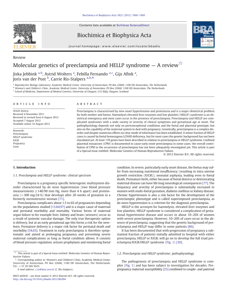
Biochimica et Biophysica Acta 1822 (2012) 1960–1969 Contents lists available at SciVerse ScienceDirect Biochimica et Biophysica Acta journal homepage: www.elsevier.com/locate/bbadis Review Molecular genetics of preeclampsia and HELLP syndrome — A review☆ Jiska Jebbink a, b, Astrid Wolters a, Febilla Fernando a, c, Gijs Afink a, Joris van der Post b, Carrie Ris-Stalpers a, b,⁎ a b c Reproductive Biology Laboratory, Academic Medical Center, University of Amsterdam, PO Box 22660, 1100 DD Amsterdam, The Netherlands Women's and Children's Clinic, Academic Medical Center, University of Amsterdam, PO Box 22660, 1100 DD Amsterdam, The Netherlands School of Medicine, Department of Medical Genetics, University of Glasgow, G12 8QQ, Glasgow, Scotland a r t i c l e i n f o Article history: Received 4 November 2011 Received in revised form 6 August 2012 Accepted 7 August 2012 Available online 16 August 2012 Keywords: Preeclampsia HELLP syndrome IUGR Pregnancy Gene a b s t r a c t Preeclampsia is characterised by new onset hypertension and proteinuria and is a major obstetrical problem for both mother and foetus. Haemolysis elevated liver enzymes and low platelets (HELLP) syndrome is an obstetrical emergency and most cases occur in the presence of preeclampsia. Preeclampsia and HELLP are complicated syndromes with a wide variety in severity of clinical symptoms and gestational age at onset. The pathophysiology depends not only on periconceptional conditions and the foetal and placental genotype, but also on the capability of the maternal system to deal with pregnancy. Genetically, preeclampsia is a complex disorder and despite numerous efforts no clear mode of inheritance has been established. A minor fraction of HELLP cases is caused by foetal homozygous LCHAD deficiency, but for most cases the genetic background has not been elucidated yet. At least 178 genes have been described in relation to preeclampsia or HELLP syndrome. Confined placental mosaicism (CPM) is documented to cause early onset preeclampsia in some cases; the overall contribution of CPM to the occurrence of preeclampsia has not been adequately investigated yet. This article is part of a Special Issue entitled: Molecular Genetics of Human Reproductive Failure. © 2012 Elsevier B.V. All rights reserved. 1. Introduction 1.1. Preeclampsia and HELLP syndrome: clinical spectrum Preeclampsia is a pregnancy specific heterogenic multisystem disorder characterised by de novo hypertension (two blood pressure measurements ≥ 140/90 mm Hg, more than 4 h apart) and proteinuria (>300 mg/24 h) that develops after 20 weeks of gestation in a formerly normotensive woman [71]. Preeclampsia complicates about 1.5 to 6% of pregnancies depending on the populations studied [13,84,97] and is a major cause of maternal and perinatal morbidity and mortality. Various forms of maternal organ failure in for example liver, kidney and brain (seizures) occur as a result of systemic vascular damage. The only true therapeutic option is delivery, but at an early gestational age this forms a risk for the newborn. Premature delivery is a major risk factor for perinatal death and morbidity [54,63]. Treatment in early preeclampsia is therefore symptomatic and aimed at prolonging pregnancy and preventing severe maternal complications as long as foetal condition allows. It consists of blood pressure regulation, seizure prophylaxis and monitoring foetal ☆ This article is part of a Special Issue entitled: Molecular Genetics of Human Reproductive Failure. ⁎ Corresponding author at: Women's and Children's Clinic, Academic Medical Center, University of Amsterdam, PO Box 22660, 1100 DD Amsterdam, The Netherlands. Tel.: + 31 20 566 5625. E-mail address: c.ris@amc.uva.nl (C. Ris-Stalpers). 0925-4439/$ – see front matter © 2012 Elsevier B.V. All rights reserved. http://dx.doi.org/10.1016/j.bbadis.2012.08.004 condition. In severe, particularly early onset disease, the foetus may suffer from increasing nutritional insufficiency (resulting in intra uterine growth restriction (IUGR)), neonatal asphyxia, leading even to foetal death. Premature birth, either because of foetal distress or deteriorating maternal disease can have life long neurological consequences [77]. The frequency and severity of preeclampsia is substantially increased in women with multi-foetal gestation, diabetes mellitus or kidney disease. Chronic hypertension is also a risk factor for the development of the preeclamptic phenotype and is called superimposed preeclampsia, as de-novo hypertension is a criterion for the diagnosis preeclampsia. HELLP is the acronym for haemolysis, elevated liver enzymes and low platelets. HELLP syndrome is considered a complication of gestational hypertensive disease and occurs in about 10–20% of women with severe preeclampsia. However, 10–20% of cases occur in the absence of preeclampsia, suggesting that the genetic background of preeclampsia and HELLP may differ in some patients [86]. It has been documented that with progression of pregnancy a substantial fraction of patients initially admitted to hospital with either preeclampsia, HELLP or IUGR, will go on to develop the full triad preeclampsia/IUGR/HELLP syndrome (Fig. 1) [30]. 1.2. Preeclampsia and HELLP syndrome: pathophysiology The pathogenesis of preeclampsia and HELLP syndrome is complex (Fig. 1) and has been subject to investigation for decades. Prepregnancy maternal susceptibility [25] combined to couple- and paternal J. Jebbink et al. / Biochimica et Biophysica Acta 1822 (2012) 1960–1969 Maternal susceptibility: • Genetic factors, incl. black ethnicity • Metabolic syndrome • Comorbidity (kidney disease, DM, hypertension, SLE) • Extremes of age Couple susceptibility: •Primiparity •KIR/HLA-C mismatch •Short cohabitual interval 1961 Paternal susceptibility: • Genetic factors • Age>45 years Black Box Insufficient placental function Release of Factor(s) X in maternal circulation endothelial dysfunction and leukocyte, complement and clotting activation time 3rd trimester 2nd trimester 1st trimester Aberrant placental implantation & Placental oxidative stress Fig. 1. Schematic representation of the events leading to the different hypertensive disorders of pregnancy. The IUGR/HELLP/Preeclampsia figure on the bottom is adapted from Ganzevoort et al. [30]. DM: diabetes mellitus. SLE: systemic lupus erythematodes. APLS: anti-phospholipid syndrome. susceptibility factors [22] contribute to the inadequate interaction between the developing placenta and the maternal endometrium. There are several key mechanisms involved that eventually lead to the clinical syndrome of preeclampsia; the immune response at the placental–maternal interface, superficial placentation with insufficient remodelling of spiral arteries, an imbalance in angiogenic factors and oxidative stress that triggers inflammation. The resulting insufficient placental function combined with release of placental factors into the maternal circulation coupled to an exaggerated maternal inflammatory response causes a generalized endothelial dysfunction and leukocyte-, complement- and clotting activation [38,69,86]. This results in the clinical syndrome of preeclampsia and HELLP syndrome (Fig. 1). 1.2.1. Immunology As the embryo expresses paternal antigens foreign to the mother's immune system active regulation of the maternal immune response at the placental–maternal interface is essential for a sustainable pregnancy [32,69]. The polymorphic HLA-C is expressed by invasive extravillous trophoblasts. HLA-C is the dominant ligand for killer immunoglobulinlike receptors (KIR) that are expressed by maternal uterine natural killer (uNK) cells. The KIR system contains two different haplotypes A and B and some KIR/HLA-C combinations are presumed to be more favourable to trophoblast-cell invasion. Due to these two polymorphic gene systems at the site of placentation, uterine NK-cell function may vary from pregnancy to pregnancy [34,62]. This immunological interface regresses in the second half of pregnancy when the villous syncytium that is devoid of HLA expression becomes dominant [11,69]. The importance of an adequate immune regulation at the placental– maternal interface is illustrated by the fact that abundant exposure to paternal antigens in seminal fluids prior to the actual pregnancy seems to prevent preeclampsia indicating some kind of ‘immunological memory’, most likely by maternal T-cells [47,72,73]. Additionally the assisted reproductive technique of oocyte donation with a high degree of antigenic dissimilarity infers an increased risk of developing pregnancy-induced hypertension [64,93]. 1.2.2. Placentation and angiogenesis Invasive cytotrophoblasts penetrate the walls of the spiral arteries where they replace maternal endothelium, stimulating remodelling of the arterial wall resulting in arterial dilatation [65]. The process of extravillous cytotrophoblasts invasion into the spiral arteries is accompanied by an ‘epithelial to endothelial’ transition involving angiogenic factors, their receptors and factors that regulate capillary function [104]. Several of these factors have been implicated in the pathogenesis of preeclampsia, like, PlGF and VEGF-A, (soluble) FLT1, TGF-beta and (soluble) Endoglin [56,96,104]. 1.2.3. Oxidative stress and inflammation The restricted invasion of cytotrophoblasts with impaired arterial remodelling of the spiral arteries results in entering of maternal blood into the intervillous space at higher pressure and faster rate. This exposes the placental villi to fluctuating oxygen concentrations 1962 J. Jebbink et al. / Biochimica et Biophysica Acta 1822 (2012) 1960–1969 2.1. Mode of inheritance, linkage analysis and the relative contribution of genetic factors. [18,68]. Oxidative stress arising from such hypoxic/re-oxygenation injuries results in widespread placental lipid and protein oxidative modifications that are pro-inflammatory. It also results in mitochondrial and endoplasmic reticulum stress, tissue apoptosis and necrosis. Additionally, oxidative stress activates NF-κB a transcription factor central to the inflammatory response and a cellular sensor of stress [2,67]. This sequence of events links oxidative stress to inflammation. The increase of necrotic trophoblast shedding due to oxidative stress mechanisms [18] may be important in the pathogenesis of preeclampsia in two ways; phagocytosis of necrotic trophoblasts results in systemic endothelial cell activation via the secretion of interleukin 6 (IL-6) [16]. Secondly, the increased amount of microparticles derived from placental syncytiotrophoblast in plasma samples from preeclamptic women are able to interact with leucocytes and monocytes and can stimulate the production of pro-inflammatory cytokines [94]. Over the years, the mode of inheritance of preeclampsia has been a matter of debate. In the 1960's through 1980's an autosomal recessive mode of inheritance was suggested, with either the maternal genotype or the foetal genotype responsible for the maternal phenotype of severe preeclampsia [17,21]. In the early 90's it was reported that homozygosity for a single recessive gene in both mother and foetus would fit 3–6% frequency of preeclampsia in the general population [58]. In the 90's a large Icelandic study concluded that either a recessive or a dominant model could fit [4]. As in preeclampsia familial clustering is not uncommon this offers the possibility to apply genome wide linkage analysis where susceptibility loci and putative candidate genes can be identified. This has been done in several studies for different populations (Table 1). Linkage analysis is an example of an approach that does not assume a specific underlying aetiology but expects a similar genotype in similarly affected patients. Linkage analysis is the ideal genome-wide method for mapping rare variants with relatively large effect sizes, but needs large families to reach statistically significant likelihood of disease (LOD) scores. Table 1 summarises the results from the PubMed search Susceptibility loci AND Preeclampsia. It shows an overview of the reported susceptibility loci and the candidate genes that could be involved in the pathogenesis of preeclampsia or HELLP syndrome found with linkage analysis. The largest sample size used to determine susceptibility loci in preeclampsia research has been 343 women (Table 1) [5] and a study this size is only able to identify genes with relatively large effects. Larger samples sizes should be able to further elucidate the genetic background of preeclampsia. Evolving insights might identify factors upstream in pathways associated with the pathophysiology. In 2001 a genome wide scan in 38 Dutch preeclampsia families revealed a locus on 10q22 subject to a parent-of-origin effect [53]. Further investigations of the families resulted in the identification of a maternally inherited mutation in exon 2 of the STOX1 gene that leads to an amino acid substitution (Y153H). This gene lies adjacent but outside the reported critical region of the originally described locus on 10q22. They described that this STOX1 amino acid variation co-segregated with the preeclampsia phenotype in seven of the eight families [95]. The epigenetic mechanism underlying this linkage was originally described as being methylation induced silencing of the paternal allele 2. Preeclampsia and HELLP syndrome: the underlying genetic basis Supplementary Table 1 lists the 178 genes, miRNAs and proteins reported in relation to preeclampsia or HELLP syndrome identified by a PubMed search Gene[title/abstract] AND preeclampsia[MESH] in the period 1989 till September 2011. The PANTHER (Protein ANalysis THrough Evolutionary Relationships) database (www.pantherdb. org) [89] was queried for the biological process relating to each gene. Of the 178 genes listed, 110 are annotated to multiple processes, 37 to only one biological process of which 22 link to a metabolic process. Of 31 genes no accompanying biological process was available, among them 12 microRNA encoding genes (MIRs). Fig. 2 is a graphical display of the relative contribution of the biological processes according to the PANTHER gene ontology database after exclusion of MIRs. The most predominant biological processes are metabolic process (m-pro), cell communication (com), immune process (imm) and response to stimuli (resp) with respectively 17, 16, 13 and 10%. The chromosomal localisation of each gene was retrieved using the NCBI data base and is schematically depicted in Fig. 3. It reveals some ‘high density regions’ with several genes implicated in preeclampsia in close vicinity to each other on chromosomes 6p, 9q, 11p and 19q. The genes displayed in this table are a mixture of genes investigated with respect to mutations or SNPs (e.g. HLA-C, FV, STOX1) and genes investigated with respect to the level of expression (e.g. FLT1, ENG). 0% 0% 0% 1% 5% 7% 2% apoptosis cell cycle 3% growth 3% 2% cell adhesion 4% transport response to stimuli 10% 17% cell communication cellular component developmental proces cellular proces system proces immune proces metabolic proces precursor metabolites 16% 13% homeostasis regulation reproduction viral reproduction 0% 5% 2% currently not annotated 8% Fig. 2. Biological processes assigned to the genes reported in relation to PE/HELLP presented in Supplementary Table 1. Genes can be allocated to one or more biological processes according to the PANTHER gene ontology database. J. Jebbink et al. / Biochimica et Biophysica Acta 1822 (2012) 1960–1969 34/35 1/2/3/ 1 42 17/7 55 43 4 5 36/37/38 9 RUFY3 6 18 7 EPAS1 45/46 19 8 11 47 56/57/ 58 ELL2/ ERAP2 (53) 62 63/64/65/ 66/67/68 77 69/70 71/72 12 13 78 73 87 91 96/97 88 79/80 SEMA3C 81/82/14 59 STOX1 98 23/24 4 27/28 29/30 10 HPS3 39 92/93/ 94/95 PAEP 11/12/13/14 5/6 38 41 119/120 121/122 108 109 123/124 110 111/112 17/18 125 113/114/ 115/116 MMP12 SART3 117 101/102 118 60/61 NOS3 50 25/26 99/100 16 89 15 83/84/85 2 9/3 10 103/104/ 105/106/ 107 90 74 48 49 20/21/22 76 1963 75 51/52/ 53/54 8 31/32/33 15/16 136 141 148 137 126 19 142/143 127/128 131 134/135 138 144/145/ 146 160 161 165 162/163 166/167 168/169 149-159 164 170/171 172 139 132 OXGR1 TNFSF13B 130 140 147 173/174 169 133 175/176 Fig. 3. Overview of chromosomal localisations of all genes reported in association with preeclampsia. Green lines represent genes retrieved by PubMed search. Red lines represent search results overlapping with susceptibility loci reported based on the analysis of chorionic villous biopsy samples from patients destined to develop preeclampsia [29]. Blue lines depict genes overlapping with susceptibility loci described in Table 1. Pink lines represent genes associated with HELLP syndrome. Confined placental mosaicism of chromosomes 13 and 16 has been reported as having an increased risk of preeclampsia (figure modified from Sasaki et al. [79]). resulting in monoallelic expression of the maternal 153H allele. We and others have shown that STOX1 is not imprinted and that both the maternal and the paternal allele are expressed in human placenta of both normotensive and preeclamptic pregnancies [37,49]. There is also no evidence for the preferential transmission of the Y153H variation from women with preeclampsia or IUGR to their offspring [8]. Despite dedicated efforts to elucidate the molecular role of STOX1 in placenta and preeclamptic placenta in particular, it remains elusive. There is an association between preeclampsia/HELLP syndrome in mothers with a child with Beckwith–Wiedemann syndrome and a mutation on the maternal CDKN1C allele [75]. CDKN1C (alias p57 KIP2) is a regulator of cell cycle control [55] and paternally imprinted [12]. Deficiency of p57kip2 expression in mice induces preeclampsia-like symptoms during pregnancy [44]. There is another example of a clear Mendelian recessive mode of inheritance in case of HELLP syndrome without preeclampsia. In pregnancies of foetuses homozygous for the Glu474Gln Long-chain 3-hydroxacyl-coenzyme A dehydrogenase (LCHAD) mutation, 77% of the heterozygous mothers develop severe pregnancy complications; acute fatty liver of pregnancy (AFLP) in 54% of mothers and HELLP syndrome in 23% [36,99]. However, LCHAD deficiency only accounts for a small percentage of HELLP cases and the genetic aetiology of HELLP syndrome remains to be unravelled [24]. Overall, there is no compelling evidence to in general regard preeclampsia as a Mendelian inherited disease and preeclampsia is currently mostly described as a ‘complex disorder’ meaning that it is believed to be associated with genetic changes combined with environmental factors [4,15]. The diversity of biological processes to which the genes in Supplementary Table 1 have been annotated and the fact that only 25% of genes are annotated to a single biological process substantiates the complex genetic background of the disease. Different approaches have been used to determine the relative contribution of genetic factors to preeclampsia. Twin studies help to distinguish between environmental and genetic influences on individual traits and behaviours. Two large twin studies on preeclampsia have been reported but the data are conflicting. In a Swedish study with 917 monozygotic and 1199 dizygotic twin pairs, the estimates of heritability and non-shared environmental effect for preeclampsia were 0.54 (95% CI 0–0.71) and 0.46 (95% CI 0.29–0.67) respectively [78]. An Australian cohort study of in total 2362 female twin pairs including only the most severe preeclamptic patients found no concordant affected twin pairs [91]. To adjust for the possible genetic or environmental contributions induced by parents, pregnancy outcomes in Swedish families over a period of 11 years were analysed using their national birth register. Information on 244,564 sib pairs with a total of 701,488 pregnancies was available. They reported that 35% of the variance in risk of preeclampsia was attributable to maternal genetic effects, 20% to foetal genetic effects (with equal contribution of maternal and paternal genetic effects), 13% to the liability of a specific couple, which is assumed to be the same in all successive pregnancies in the same couple, less than 1% to shared sib environment, and 32% to undetermined factors [19]. 1964 Table 1 Reported susceptibility loci for preeclampsia. Candidate genes in bold were confirmed by Founds S.A. et al. [28] by comparing susceptibility loci with gene expression data obtained from microarray analysis of chorionic villous sampling (CVS) from women whose pregnancies were complicated by preeclampsia. Candidate gene LOD-score/ NPL-score Technique Population Nr of cases 4q RUFY3 2.9L Genome wide linkage study Australian 15 pedigrees 2p13 EPAS 4.7L Genome wide screen Icelandic 343 Genome wide linkage study Australian 26 families Medium density genome scan Australian/New Zealand 121 women Genome wide linkage study Dutch 67 sibpair families Finnish 305 Finnish 15 families L 7q36 NOS3/SEMA3C 2.143 2p12 11q23-q24 2q23 12q 22q13.1 10q22.1 11q13 EPAS MMP12 FN1/ACVR2 SART3 STOX1 2.58L 2.02L 3.43L 1.99L 2.41L 2.38L 2p12-p13 EPAS P = 0.04 2p25 9p13 4q32 3q11.1-21.2 7q34-7q36.3 9q34.1-9q34.3 2q37.1-2q37.3 5q 13q EPAS TLR2 HPS3 SEMA3C PAEP FN1 ELL2/ERAP OXGR1 3.77 3.74 3.13 N Genome wide screen Clinical definition PE N HEGESMAe 3.12L 3.10L Medium density genome scan Australian/New Zealand 34 families L = LOD-score (LOD scores of 0.59, 1.17 and 2.07 correspond to significance levels of b0.05, b0.01 and b0.001 respectively), respectively) [51]. All clinical features stated appeared after 20 weeks of gestation. a SPE = severe preeclampsia. b MPE = mild preeclampsia. c BP = bloodpressure. d GH = gestational hypertension. e HEGESMA = heterogeneity-based genome search meta-analysis. Blood pressure/laboratory results Proteinuria SPEa: BPb > 140/90 mm Hg MPEc: BP> 140/90 without proteinuria General criteria: GHd, PE or E Strict criteria: PE or E MPE: BP > 140/90 mm Hg SPE: BP > 140/90 mm Hg E: all above mentioned with seizures MPE: BP > 140/90 mm Hg SPE: BP > 140/90 mm Hg E: all above mentioned with seizures MPE: de novo hypertension PE: BP> 140/90 mm Hg E: above mentioned complicated with seizures HELLP: LDHc > 600 IU/l and ASATd and ALATe >70 IU/l and b100 platelets*10^9/l PE: BP> 140/90 mm Hg >0.3 g/l protein in 24 h MPE: BP > 140/90 mm Hg SPE: BP > 160/110 mm Hg E : all above mentioned with seizures MPE: BP > 140/90 mm Hg SPE: BP > 140/90 mm Hg E: all above mentioned with seizures N MPE: BP > 140/90 mm Hg SPE: BP > 140/90 mm Hg PE: all above mentioned with seizures N Author Harrison et al. 1997 Arngrimsson et al. 1999 MPE: no proteinuria SPE: >0.3 g/l protein in 24 h Guo et al.1999 MPE: no proteinuria SPE: >0.3 g/l protein in 24 h Moses et al. 2000 Fitzpatrick et al. 2004 MPE: no proteinuria PE: >0.3 g/l proteinuria Lachmeijer et al. 2001 PE: new onset proteinuria of >300 mg MPE: no proteinuria SPE: >2 g/l protein in 24 h Laasanen et al. 2003 MPE: no proteinuria SPE: >0.3 g/l protein in 24 h Zintzaras et al. 2006 MPE: no proteinuria SPE: >0.3 g/l protein in 24 h Johnson et al. 2007 Laivuori et al. 2003 = NPL-score (NPL scores of 1.65, 2.33 and 3.09 correspond to significance levels of b0.05, b0.01 and b0.001 J. Jebbink et al. / Biochimica et Biophysica Acta 1822 (2012) 1960–1969 Locus J. Jebbink et al. / Biochimica et Biophysica Acta 1822 (2012) 1960–1969 The more severe forms of preeclampsia seem to harbour a stronger genetic component [85]. In conclusion: most studies report that the genetic contribution to the development of preeclampsia is around 50% implying that gene–environment interactions play a role. Both excess homocysteine and dietary deficiencies of folate and vitamins B6 and B12 have been implicated in the pathogenesis of preeclampsia, although results are not unequivocal [6,26,45,59]. The proposed mechanism of hyperhomocysteinemia-induced pre-eclampsia is that homocysteine can accumulate through either increased dietary methionine or a deficiency of B vitamins and folate. Excess homocysteine can then be converted to S-adenosyl homocysteine (SAH) through the enzyme SAH hydrolase. High levels of SAH can inhibit catecholO-methyltransferase (COMT), an enzyme that metabolizes estradiols. Decreased COMT activity can deplete levels of 2-ME, a metabolite of COMT capable of regulating HIF-1a levels. COMT deficiency is associated with preeclampsia in mice [43]. Although the supplementation of vitamins C and E as anti-oxidants to reduce oxidative stress and prevent preeclampsia seemed promising at first [14], randomized trials do not support a role for vitamins C and E in preventing preeclampsia [20]. One trial even showed that vitamins C and E increase the risk of foetal loss or perinatal death [102]. Although smoking during pregnancy may lead to many adverse effects such as foetal growth restriction, placental abruption, stillbirth, and preterm labour, smoking is the only environmental exposure known to consistently reduce the risk of preeclampsia and gestational hypertension [98]. The protective mechanism is still under research. 1965 Recently, the presence of placental trisomy by comparative genomic hybridisation was investigated in 43 IUGR placentas (of which 25 were associated with preeclampsia), 18 preeclamptic placentas and 11 placentas with abnormal maternal serum findings in relation to trisomy 21 screening. Of these 72 placentas analysed, 6 placentas had placental trisomy. Two out of six cases with placental trisomy had onset of preeclampsia before the 34th week of gestation. In none of the 85 control placentas placental CPM was observed [74]. Fig. 3 depicts that only a few genes previously investigated in relation to preeclampsia are located on chromosomes 13 and 16. Most genes associated with preeclampsia, listed in Supplementary Table 1 located on chromosome 13 or 16 were indeed identified based on differential expression. Coagulation factor 7(F7), located on 13q34, was identified based on investigations on preeclampsia in women who delivered a trisomy 13 [92]. 2.3. Identification of candidate genes based on pathophysiology Many investigators apply a hypothesis driven approach where they investigate associations between the disease and changes in candidate genes implied in the pathogenesis. 2.3.1. Immunology Some KIR/HLA-C combinations appear unfavourable to trophoblastcell invasion [61]. Mothers with an AA killer immunoglobulin-like receptors (KIR) genotype and a foetus with a paternal HLA-C2 are at greatly increased risk of a preeclamptic pregnancy [34,69]. Additionally, genetic susceptibility of HLA-DR4 with preeclampsia has been described [48]. 2.2. Confined placental mosaicism in relation to preeclampsia With the exception of those involving chromosomes 13, 18 or 21 all trisomic pregnancies tend to undergo spontaneous abortions. So when an ongoing pregnancy with for instance a trisomy 3 is diagnosed prenatally it is considered to be a confined placental mosaicism (CPM). The incidence of CPM in chorionic villous samples is 1–2% [74]. Trisomy in placenta can affect cytotrophoblast differentiation, which is a prerequisite for proper placentation [100]. Trisomy mainly occurs due to non-disjunction events [41]. Based on the tissue of confinement of trisomy, CPM is differentiated into three types; CPM confined to cytotrophoblasts (type I), confined to mesenchymal core (type II) and CPM present in both cytotrophoblasts and mesenchymal core (type III) [90]. Type I is the most commonly occurring CPM subtype and no adverse pregnancy outcomes have been reported. CPM type III occurs less frequently, but is commonly associated with a poor pregnancy outcome [74]. Type III CPM usually has an early meiotic origin of the error (non-disjunction) giving rise to a trisomic embryo; subsequent rescue of trisomy in progenitor cells of foetus leaves the placenta almost entirely trisomic (Fig. 4). Some studies have reported that presence of a type III placental trisomy can cause IUGR [74,90] or preeclampsia [3,9,74]. Especially CPM of trisomy 13 has been reported as having an increased risk of preeclampsia [7,9,33,50]. This might relate to the fact that the FLT1 gene is localized on 13q12.3. sFLT1 protein levels in maternal serum of pregnancies with a trisomy 13 confined to the placenta, were 35% higher compared to normal pregnancies [7]. Additionally, in nonpregnant rats the administration of sFLT1 evokes preeclampsia like symptoms [60]. All evidence points to a gene-dosage effect of sFLT1 in preeclampsia. Placental trisomy 16 has also been reported in association with preeclampsia [10,42] with a 3–4 times increased risk of preeclampsia compared to a control population [103]. The occurrence of preeclampsia in 25 prenatally diagnosed mosaic trisomy 16 pregnancies was investigated and higher levels of trisomy were observed in all placental lineages of preeclamptic cases when compared to the non-preeclamptic cases. Uniparental disomy (UPD) did not seem to influence the risk of pre-eclampsia in this study [103]. 2.3.2. Placentation and angiogenesis The vascular endothelial growth factor (VEGF) ligands and their receptors play an essential role in both normal and pathological functioning of the endothelium [87]. VEGF receptor 1 (VEGFR1) also known as FLT1 (Fms-like tyrosine kinase 1) is a transmembrane tyrosine kinase type receptor with multiple ligands (Placental growth factor (PlGF), VEGF-A and VEGF-B). Ultimately activation of these receptors plays a key role in angiogenesis [76]. Apart from the transmembrane form of VEGFR1, there is a soluble form lacking the transmembrane domain. This soluble truncated version of VEGF receptor 1 (also known as sFLT1) is markedly elevated in the circulation of preeclamptic women [60]. The current concept of the role of sFLT1 in preeclampsia is that it traps its ligands VEGF and PlGF, thereby lowering free circulating levels of these factors below a critical threshold. sFLT1 mRNA is generated by alternative splicing of the FLT1 gene. The discovery of additional alternative spliced FLT1 transcripts encoding novel soluble (s)FLT1 protein isoforms [46,80,88] complicates both the predictive value and functional implications of sFLT1 in preeclampsia. Placenta has by far the highest FLT1 mRNA expression level compared to other tissues and expression is directly up-regulated by hypoxia via a hypoxia-inducible enhancer element in the FLT1 gene promoter [31]. Over 80% of placental transcripts correspond to sFLT1_v2. Placental FLT1 transcript levels are increased not only in preeclampsia but also in normotensive pregnancy with a small for gestational age foetus. This may indicate a common pathway involved in the development of both conditions [39]. Injection of sFLT1 expressing adenovirus in rats results in increased blood pressure and proteinuria, but this is pregnancy independent [60]. A mouse model with placenta-specific sFLT1 expression demonstrates hypertension and proteinuria in pregnancy that resolve after delivery [52]. Endoglin is an auxiliary cell surface receptor for the transforming growth factor beta 1(TGF-β1) and TGF-β3 that are potent inhibitors of trophoblast differentiation and migration. Soluble endoglin, the product of proteolytic cleavage of endoglin, inhibits the action of TGF‐β1 and TGF-β3. The expression of ENG and the production of soluble endoglin is upregulated in preeclamptic placenta. Elevated serum soluble endoglin levels correlate with disease severity. Soluble endoglin is able to produce increased vascular permeability and hypertension in 1966 J. Jebbink et al. / Biochimica et Biophysica Acta 1822 (2012) 1960–1969 Fig. 4. CPM by meiotic non-disjunction and trisomy rescue in the foetus: a meiotic non-disjunction event in one of the parental gametes gives rise to a diploid gamete which subsequently fuses with a normal gamete resulting in a trisomic embryo. In a later stage during post-zygotic divisions the progenitor cells of the foetus undergo trisomy rescue and lose the extra chromosome. The foetus becomes diploid and as a result the trisomy is confined to the placenta. rats in vivo. Co-administration of sEng and sFlt1 expressing adenovirus in rats results even in a more severe preeclamptic phenotype combined with HELLP [96]. The identity of the protein responsible for the increased cleavage of endoglin and the production of the soluble form has recently been attributed to MMP-14 [40]. Soluble endoglin is upregulated in preeclampsia in a pattern similar to sFLT1 [83]. 2.3.3. Oxidative stress and inflammation In placenta, COMT metabolizes estradiols to 2-methoxy-estradiol, an estradiol metabolite that destabilizes hypoxia-inducible factor (HIF)-1a. HIF proteins mediate the effects of hypoxia on gene expression by up regulating transcription of target genes, including FLT1 with preference for the alternative splice product sFLT1. This role of COMT in maintaining oxygen balance suggests that COMT might somehow be involved in the pathogenesis of pre-eclampsia through an altered response to oxidative stress [82]. Recently it has been shown that homozygosity for the variant allele of the maternal COMT gene may increase susceptibility to preeclampsia [57]. This is further supported by the fact that compared to wild type mice, comt−/− mice have increased blood pressures, higher levels of proteinuria, smaller offspring and smaller placenta's [43]. COMT has also been shown to interact with methylenetetrahydrofolate reductase (MTHFR), which modulates the availability of S-adenosylmethionine (SAM), a COMT cofactor. Variations in both foetal and maternal MTHFR have been associated with preeclampsia [35]. In addition some mothers and foetus are more susceptible to oxidative stress compared to others due to impaired function in scavenger molecules like glutathione S-transferases (GTS) P1, M1 and T1, epoxide hydrolase (EPHX) and cytochrome P4501A1 (CYP1A1) [105]. Inflammatory cytokines like TNF-alpha, IL-6 and IL-10 have been implicated to contribute to the pathological inflammation process seen in preeclampsia. In 2011 a systematic review of preeclampsia in relation to polymorphisms and circulating concentrations of these cytokines reported that maternal TNF-a-308G/A, IL-6 174G/C and IL-10-1082A/G polymorphisms were not associated with preeclampsia [101]. On the other hand, maternal serum concentrations of all three cytokines were significantly higher in preeclampsia patients versus controls. These findings strengthen the clinical evidence that preeclampsia is accompanied by an exaggerated inflammatory response, but do not support TNF-α-308G/A, IL-6-174G/C, and IL-10-1082A/G as candidate susceptibility loci in preeclampsia [101]. In 2006 it was reported that 7 genes (AGT, (angiotensinogen), AGTR1 and AGTR2 (the angiotensin receptors), FV (coagulation factor v), MTHFR (methylenetetrahydrofolate reductase), NOS3 (nitric oxide synthase 3) and TNF-α (tumour necrosis factor-alpha)) dominate 70% of literature regarding the genetics of preeclampsia [15]. The interest in these genes can be explained by their involvement in different underlying aetiologies relevant to the pathogenesis of preeclampsia. Angiotensinogen and the angiotensin receptors are part of the renin–angiotensin system that regulates blood pressure [23]. Transgenic mice overexpressing human renin and angiotensinogen develop superimposed preeclampsia [27]. Factor V is part of the blood coagulation pathway and severe and early-onset preeclampsia is significantly associated with inherited thrombophilia [66]. MTHFR plays a role in homocysteine metabolism [35]. NOS3 (also known as endothelial NOS) is important in vasodilation required to accommodate the increased circulating volume during pregnancy without a rise in blood pressure [81] and TNF-alpha is an important apoptosis inducer [70]. The GOPEC consortium analysed these 7 candidate genes reported as conferring susceptibility to preeclampsia in 627 UK families with preeclampsia (including 398 maternal triads and 536 foetal triads). Using the transmission disequilibrium test, no genotype risk ratio achieved the pre specified criteria for statistical significance [1]. J. Jebbink et al. / Biochimica et Biophysica Acta 1822 (2012) 1960–1969 3. In conclusion Since the discovery of DNA in the 1960's numerous efforts have been made to elucidate the genetic background of preeclampsia; alas without comprehensive results. Currently, there is no established genotype–phenotype relation for preeclampsia. A pitfall in defining the mode of inheritance has been the spectrum of different subtypes of women who all meet the ISSHP criteria [71] of de novo hypertension and proteinuria. The most likely reason is that we are dealing with a complex disorder; preeclampsia and HELLP are complicated syndromes with a very wide range of clinical symptoms depending not only on periconceptional conditions and the foetal and placental genotype, but also on the capability of the maternal system to deal with the reproductive challenge that pregnancy is. For HELLP syndrome one example of a genotype– phenotype correlation exist; heterozygotic women carrying a LCHAD deficient foetus have a 77% risk of AFLP or HELLP syndrome. At present there are very few studies that have investigated the presence of placental aneuploidy in preeclamptic cases and therefore it is difficult to give an appropriate risk estimate based on the available data. Prenatally diagnosed trisomy 13 and trisomy 16 have an increased risk of pre-eclampsia when compared to the other trisomies and should be provided better obstetrical supervision. Data from micro-array and SAGE analysis report genes with altered expression in preeclampsia/HELLP placenta. Theoretically the cause for this altered expression can be within the causative gene, in an upstream factor influencing transcription, or in a downstream factor influencing mRNA stability. With the exemption of HIF-1a the data coming from the diverse molecular studies have not been fully integrated yet. 4. Current challenges and future perspectives The ISSHP criteria for preeclampsia are generally accepted across the world. The next step should be to reach international consensus on criteria for the different sub-types of preeclampsia varying from mild hypertension and some proteinuria at term to severe hypertension, proteinuria, eclampsia and additional laboratory abnormalities at an early gestational age. Factors like predisposing maternal factors, a positive family history of preeclampsia, early onset disease and the combination with either IUGR and/or HELLP syndrome should be taken into account to investigate causative factors in homogenous subgroups of women who all meet the ISSHP criteria of preeclampsia. International collaboration is essential to reach adequate power to detect the molecular cause of specific sub-types. Basic research based on pathophysiological mechanisms will be essential to provide insights and tell us whether previous findings are cause or consequence. More research is needed to determine the true incidence of CPM in relation to preeclampsia. Given undisputed involvement of sFLT1 in the aetiology of preeclampsia, the location of the FLT1 gene on chromosome 13 and the consistent evidence of raised levels of sFLT1 in preeclamptic women this is worth pursuing. Ultimately, the firm establishment of the genetic basis of preeclampsia and HELLP syndrome will provide a rational basis for the development of further prognostic and therapeutic targets. References [1] GOPEC Consortium, Disentangeling fetal and maternal susceptibility for preeclampsia: a British multicentercandidate-gene study, Am. J. Hum. Genet. 77 (2005) 127–131. [2] K.S. Ahn, B.B. Aggarwal, Transcription factor NF-kappaB: a sensor for smoke and stress signals, Ann. N. Y. Acad. Sci. 1056 (2005) 218–233. [3] A. Amiel, N. Bouaron, D. Kidron, R. Sharony, E. Gaber, M.D. Fejgin, CGH in the detection of confined placental mosaicism (CPM) in placentas of abnormal pregnancies, Prenat. Diagn. 22 (2002) 752–758. [4] R. Arngrímsson, S. Björnsson, R.T. Geirsson, H. Björnsson, J.J. Walker, G. Snaedal, Genetic and familial predisposition to eclampsia and pre-eclampsia in a defined population, Br. J. Obstet. Gynaecol. 97 (1990) 762–769. 1967 [5] R. Arngrímsson, S. Sigurõardóttir, M.L. Frigge, R.I. Bjarnadóttir, T. Jóhnson, H. Stefánsson, A. Baldursdóttir, A.S. Einarsdóttir, B. Palsson, S. Snorradóttir, A.M. Lachmeijer, D. Nicolae, A. Kong, B.T. Bragason, J.R. Gulcher, R.T. Geirsson, K. Stefánsson, A genome-wide scan reveals a maternal susceptibility locus for pre-eclampsia on chromosome 2p13, Hum. Mol. Genet. 8 (1999) 1799–1805. [6] A. Baksu, M. Taskin, N. Goker, B. Baksu, A. Uluocak, Plasma homocysteine in late pregnancies complicated with preeclampsia and in newborns, Am. J. Perinatol. 23 (2006) 31–35. [7] Y. Bdolah, G.E. Palomaki, Y. Yaron, T. Bdolah-Abram, M. Goldman, R.J. Levine, B.P. Sachs, J.E. Haddow, S.A. Karumanchi, Circulating angiogenic proteins in trisomy 13, Am. J. Obstet. Gynecol. 194 (2006) 239–245. [8] A.L. Berends, A.M. Bertoli-Avella, C.J. de Groot, C.M. van Duijn, B.A. Oostra, E.A. Steegers, STOX1 gene in pre-eclampsia and intrauterine growth restriction, BJOG. 114 (2007) 1163–1167. [9] P.A. Boyd, R.H. Lindenbaum, C. Redman, Pre-eclampsia and trisomy 13: a possible association, Lancet 2 (1987) 425–427. [10] H. Brandenburg, F.J. Los, P. in 't Veld, Clinical significance of placenta-confined nonmosaic trisomy 16, Am. J. Obstet. Gynecol. 174 (1996) 1663–1664. [11] I.A. Brosens, W.B. Robertson, H.G. Dixon, The role of the spiral arteries in the pathogenesis of preeclampsia, Obstet. Gynecol. Annu. 1 (1972) 177–191. [12] D.H. Castrillon, D. Sun, S. Weremowicz, R.A. Fisher, C.P. Crum, D.R. Genest, Discrimination of complete hydatidiform mole from its mimics by immunohistochemistry of the paternally imprinted gene product p57KIP2, Am. J. Surg. Pathol. 25 (2001) 1225–1230. [13] A.B. Caughey, N.E. Stotland, A.E. Washington, G.J. Escobar, Maternal ethnicity, paternal ethnicity, and parental ethnic discordance: predictors of preeclampsia, Obstet. Gynecol. 106 (2005) 156–161. [14] L.C. Chappell, P.T. Seed, A.L. Briley, F.J. Kelly, R. Lee, B.J. Hunt, K. Parmar, S.J. Bewley, A.H. Shennan, P.J. Steer, L. Poston, Effect of antioxidants on the occurrence of pre-eclampsia in women at increased risk: a randomised trial, Lancet 354 (1999) 810–816. [15] S. Chappell, L. Morgan, Searching for genetic clues to the causes of pre-eclampsia, Clin. Sci. (Lond.) 110 (2006) 443–458. [16] Q. Chen, P. Stone, L.M. Ching, L. Chamley, A role for interleukin-6 in spreading endothelial cell activation after phagocytosis of necrotic trophoblastic material: implications for the pathogenesis of pre-eclampsia, J. Pathol. 217 (2009) 122–130. [17] L.C. Chesley, D.W. Cooper, Genetics of hypertension in pregnancy: possible single gene control of pre-eclampsia and eclampsia in the descendants of eclamptic women, Br. J. Obstet. Gynaecol. 93 (1986) 898–908. [18] T. Cindrova-Davies, Gabor Than Award Lecture 2008: pre-eclampsia - from placental oxidative stress to maternal endothelial dysfunction, Placenta 30 (Suppl. A) (2009). [19] S. Cnattingius, M. Reilly, Y. Pawitan, P. Lichtenstein, Maternal and fetal genetic factors account for most of familial aggregation of preeclampsia: a population-based Swedish cohort study, Am. J. Med. Genet. A 130A (2004) 365–371. [20] A. Conde-Agudelo, R. Romero, J.P. Kusanovic, S.S. Hassan, Supplementation with vitamins C and E during pregnancy for the prevention of preeclampsia and other adverse maternal and perinatal outcomes: a systematic review and metaanalysis, Am. J. Obstet. Gynecol. 204 (2011) 503–512. [21] D.W. Cooper, W.A. Liston, Genetic control of severe pre-eclampsia, J. Med. Genet. 16 (1979) 409–416. [22] G. Dekker, P.Y. Robillard, C. Roberts, The etiology of preeclampsia: the role of the father, J. Reprod. Immunol. 89 (2011) 126–132. [23] C. Delles, M.W. McBride, D. Graham, S. Padmanabhan, A.F. Dominiczak, Genetics of hypertension: from experimental animals to humans, Biochim. Biophys. Acta 1802 (2010) 1299–1308. [24] M.E. den Boer, L. Ijlst, F.A. Wijburg, W. Oostheim, M.A. van Werkhoven, M.G. van Pampus, H.S. Heymans, R.J. Wanders, Heterozygosity for the common LCHAD mutation (1528g>C) is not a major cause of HELLP syndrome and the prevalence of the mutation in the Dutch population is low, Pediatr. Res. 48 (2000) 151–154. [25] K. Duckitt, D. Harrington, Risk factors for pre-eclampsia at antenatal booking: systematic review of controlled studies, BMJ 330 (2005) 565. [26] S. Falcao, S. Bisotto, J. Gutkowska, J.L. Lavoie, Hyperhomocysteinemia is not sufficient to cause preeclampsia in an animal model: the importance of folate intake, Am. J. Obstet. Gynecol. 200 (2009) 198.e.1–5. [27] S. Falcao, E. Stoyanova, G. Cloutier, R.L. Maurice, J. Gutkowska, J.L. Lavoie, Mice overexpressing both human angiotensinogen and human renin as a model of superimposed preeclampsia on chronic hypertension, Hypertension 54 (2009) 1401–1407. [28] S.A. Founds, Y.P. Conley, J.F. Lyons-Weiler, A. Jeyabalan, W.A. Hogge, K.P. Conrad, Altered global gene expression in first trimester placentas of women destined to develop preeclampsia, Placenta 30 (2009) 15–24. [29] S.A. Founds, L.A. Terhorst, K.P. Conrad, W.A. Hogge, A. Jeyabalan, Y.P. Conley, Gene expression in first trimester preeclampsia placenta, Biol. Res. Nurs. 13 (2011) 134–139. [30] W. Ganzevoort, A. Rep, G.J. Bonsel, J.I. de Vries, H. Wolf, Dynamics and incidence patterns of maternal complications in early-onset hypertension of pregnancy, BJOG. 114 (2007) 741–750. [31] H.P. Gerber, F. Condorelli, J. Park, N. Ferrara, Differential transcriptional regulation of the two vascular endothelial growth factor receptor genes. Flt-1, but not Flk-1/KDR, is up-regulated by hypoxia, J. Biol. Chem. 272 (1997) 23659–23667. [32] G. Girardi, Z. Prohászka, R. Bulla, F. Tedesco, S. Scherjon, Complement activation in animal and human pregnancies as a model for immunological recognition, Mol. Immunol. 48 (2011) 1621–1630. 1968 J. Jebbink et al. / Biochimica et Biophysica Acta 1822 (2012) 1960–1969 [33] R. Heydanus, P. Defoort, M. Dhont, Pre-eclampsia and trisomy 13, Eur. J. Obstet. Gynecol. Reprod. Biol. 60 (1995) 201–202. [34] S.E. Hiby, J.J. Walker, K.M. O'shaughnessy, C.W. Redman, M. Carrington, J. Trowsdale, A. Moffett, Combinations of maternal KIR and fetal HLA-C genes influence the risk of preeclampsia and reproductive success, J. Exp. Med. 200 (2004) 957–965. [35] L.D. Hill, T.P. York, J.P. Kusanovic, R. Gomez, L.J. Eaves, R. Romero, J.F. Strauss III, Epistasis between COMT and MTHFR in maternal-fetal dyads increases risk for preeclampsia, PLoS One 6 (2011) e16681. [36] J.A. Ibdah, M.J. Bennett, P. Rinaldo, Y. Zhao, B. Gibson, H.F. Sims, A.W. Strauss, A fetal fatty-acid oxidation disorder as a cause of liver disease in pregnant women, N. Engl. J. Med. 340 (1999) 1723–1731. [37] I. Iglesias-Platas, D. Monk, J. Jebbink, M. Buimer, K. Boer, F. van der Hills, S. Apostolidou, C. Ris-Stalpers, P. Stanier, G.E. Moore, STOX1 is not imprinted and is not likely to be involved in preeclampsia, Nat. Genet. 39 (2007) 279–280. [38] J.L. James, G.S. Whitley, J.E. Cartwright, Pre-eclampsia: fitting together the placental, immune and cardiovascular pieces, J. Pathol. 221 (2010) 363–378. [39] J. Jebbink, R. Keijser, G. Veenboer, J. van der Post, C. Ris-Stalpers, G. Afink, Expression of placental FLT1 transcript variants relates to both gestational hypertensive disease and fetal growth, Hypertension 58 (2011) 70–76. [40] T.J. Kaitu'u-Lino, K.R. Palmer, C.L. Whitehead, E. Williams, M. Lappas, S. Tong, MMP-14 is expressed in preeclamptic placentas and mediates release of soluble endoglin, Am. J. Pathol. 180 (2012) 888–894. [41] D.K. Kalousek, Pathogenesis of chromosomal mosaicism and its effect on early human development, Am. J. Med. Genet. 91 (2000) 39–45. [42] D.K. Kalousek, S. Langlois, I. Barrett, I. Yam, D.R. Wilson, P.N. Howard-Peebles, M.P. Johnson, E. Giorgiutti, Uniparental disomy for chromosome 16 in humans, Am. J. Hum. Genet. 52 (1993) 8–16. [43] K. Kanasaki, K. Palmsten, H. Sugimoto, S. Ahmad, Y. Hamano, L. Xie, S. Parry, H.G. Augustin, V.H. Gattone, J. Folkman, J.F. Strauss, R. Kalluri, Deficiency in catechol-O-methyltransferase and 2-methoxyoestradiol is associated with preeclampsia, Nature 453 (2008) 1117–1121. [44] N. Kanayama, K. Takahashi, T. Matsuura, M. Sugimura, T. Kobayashi, N. Moniwa, M. Tomita, K. Nakayama, Deficiency in p57Kip2 expression induces preeclampsialike symptoms in mice, Mol. Hum. Reprod. 8 (2002) 1129–1135. [45] S.E. Kassab, M.F. Abu-Hijleh, H.B. Al-Shaikh, D.S. Nagalla, Hyperhomocysteinemia in pregnant rats: effects on arterial pressure, kidneys and fetal growth, Eur. J. Obstet. Gynecol. Reprod. Biol. 122 (2005) 177–181. [46] R.L. Kendall, K.A. Thomas, Inhibition of vascular endothelial cell growth factor activity by an endogenously encoded soluble receptor, Proc. Natl. Acad. Sci. U. S. A. 90 (1993) 10705–10709. [47] E.M. Kho, L.M. McCowan, R.A. North, C.T. Roberts, E. Chan, M.A. Black, R.S. Taylor, G.A. Dekker, Duration of sexual relationship and its effect on preeclampsia and small for gestational age perinatal outcome, J. Reprod. Immunol. 82 (2009) 66–73. [48] D.C. Kilpatrick, W.A. Liston, F. Gibson, J. Livingstone, Association between susceptibility to pre-eclampsia within families and HLA DR4, Lancet 2 (1989) 1063–1065. [49] K. Kivinen, H. Peterson, L. Hiltunen, H. Laivuori, S. Heino, I. Tiala, S. Knuutila, V. Rasi, J. Kere, Evaluation of STOX1 as a preeclampsia candidate gene in a population-wide sample, Eur. J. Hum. Genet. 15 (2007) 494–497. [50] M.P. Koster, P. Stoutenbeek, G.H. Visser, P.C. Schielen, Trisomy 18 and 13 screening: consequences for the Dutch Down syndrome screening programme, Prenat. Diagn. 30 (2010) 287–289. [51] L. Kruglyak, M.J. Daly, M.P. Reeve-Daly, E.S. Lander, Parametric and nonparametric linkage analysis: a unified multipoint approach, Am. J. Hum. Genet. 58 (1996) 1347–1363. [52] K. Kumasawa, M. Ikawa, H. Kidoya, H. Hasuwa, T. Saito-Fujita, Y. Morioka, N. Takakura, T. Kimura, M. Okabe, Pravastatin induces placental growth factor (PGF) and ameliorates preeclampsia in a mouse model, Proc. Natl. Acad. Sci. U. S. A. 108 (2011) 1451–1455. [53] A.M. Lachmeijer, R. Arngrímsson, E.J. Bastiaans, M.L. Frigge, G. Pals, S. Sigurõardóttir, H. Stefánsson, B. Pálsson, D. Nicolae, A. Kong, J.G. Aarnoudse, J.R. Gulcher, G.A. Dekker, L.P. ten Kate, K. Stefánsson, A genome-wide scan for preeclampsia in the Netherlands, Eur. J. Hum. Genet. 9 (2001) 758–764. [54] B. Larroque, G. Bréart, M. Kaminski, M. Dehan, M. André, A. Burguet, H. Grandjean, B. Ledésert, C. Lévêque, F. Maillard, J. Matis, J.C. Rozé, P. Truffert, Survival of very preterm infants: Epipage, a population based cohort study, Arch. Dis. Child. Fetal Neonatal Ed. 89 (2004) F139–F144. [55] M.H. Lee, I. Reynisdottir, J. Massague, Cloning of p57KIP2, a cyclin-dependent kinase inhibitor with unique domain structure and tissue distribution, Genes Dev. 9 (1995) 639–649. [56] R.J. Levine, C. Lam, C. Qian, K.F. Yu, S.E. Maynard, B.P. Sachs, B.M. Sibai, F.H. Epstein, R. Romero, R. Thadhani, S.A. Karumanchi, Soluble endoglin and other circulating antiangiogenic factors in preeclampsia, N. Engl. J. Med. 355 (2006) 992–1005. [57] J.H. Lim, S.Y. Kim, d.J. Kim, S.Y. Park, H.W. Han, J.Y. Han, S.W. Lee, J.H. Yang, H.M. Ryu, Genetic polymorphism of catechol-O-methyltransferase and cytochrome P450c17alpha in preeclampsia, Pharmacogenet. Genomics 20 (2010) 605–610. [58] W.A. Liston, D.C. Kilpatrick, Is genetic susceptibility to pre-eclampsia conferred by homozygosity for the same single recessive gene in mother and fetus? Br. J. Obstet. Gynaecol. 98 (1991) 1079–1086. [59] G. Maruotti, B.A. Del, A.N. Amato, L. Lombardi, A.M. Fulgeri, F. Pietropaolo, Preeclampsia and high serum levels of homocysteine, Minerva Ginecol. 57 (2005) 165–170. [60] S.E. Maynard, J.Y. Min, J. Merchan, K.H. Lim, J. Li, S. Mondal, T.A. Libermann, J.P. Morgan, F.W. Sellke, I.E. Stillman, F.H. Epstein, V.P. Sukhatme, S.A. Karumanchi, Excess placental soluble fms-like tyrosine kinase 1 (sFlt1) may contribute to [61] [62] [63] [64] [65] [66] [67] [68] [69] [70] [71] [72] [73] [74] [75] [76] [77] [78] [79] [80] [81] [82] [83] [84] [85] [86] [87] [88] [89] [90] endothelial dysfunction, hypertension, and proteinuria in preeclampsia, J. Clin. Invest. 111 (2003) 649–658. A. Moffett, S.E. Hiby, How Does the maternal immune system contribute to the development of pre-eclampsia? Placenta 28 (Suppl. A) (2007) S51–S56. A. Moffett, C. Loke, Immunology of placentation in eutherian mammals, Nat. Rev. Immunol. 6 (2006) 584–594. D. Moster, R.T. Lie, T. Markestad, Long-term medical and social consequences of preterm birth, N. Engl. J. Med. 359 (2008) 262–273. U. Pecks, N. Maass, J. Neulen, Oocyte donation: a risk factor for pregnancy-induced hypertension: a meta-analysis and case series, Dtsch. Arztebl. Int. 108 (2011) 23–31. R. Pijnenborg, J. Anthony, D.A. Davey, A. Rees, A. Tiltman, L. Vercruysse, A. van Assche, Placental bed spiral arteries in the hypertensive disorders of pregnancy, Br. J. Obstet. Gynaecol. 98 (1991) 648–655. W. Rath, Pre-eclampsia and inherited thrombophilia: a reappraisal, Semin. Thromb. Hemost. 37 (2011) 118–124. C.W. Redman, Preeclampsia: a multi-stress disorder, Rev. Med. Interne 32 (Suppl. 1) (2011) S41–S44. C.W. Redman, I.L. Sargent, Latest advances in understanding preeclampsia, Science 308 (2005) 1592–1594. C.W. Redman, I.L. Sargent, Immunology of pre-eclampsia, Am. J. Reprod. Immunol. 63 (2010) 534–543. F. Reister, H.G. Frank, J.C. Kingdom, W. Heyl, P. Kaufmann, W. Rath, B. Huppertz, Macrophage-induced apoptosis limits endovascular trophoblast invasion in the uterine wall of preeclamptic women, Lab. Invest. 81 (2001) 1143–1152. J.M. Roberts, G. Pearson, J. Cutler, M. Lindheimer, Summary of the NHLBI Working Group on Research on Hypertension During Pregnancy, Hypertension 41 (2003) 437–445. P.Y. Robillard, G. Dekker, G. Chaouat, T.C. Hulsey, A. Saftlas, Epidemiological studies on primipaternity and immunology in preeclampsia—a statement after twelve years of workshops, J. Reprod. Immunol. 89 (2011) 104–117. P.Y. Robillard, T.C. Hulsey, Association of pregnancy-induced-hypertension, pre-eclampsia, and eclampsia with duration of sexual cohabitation before conception, Lancet 347 (1996) 619. W.P. Robinson, M.S. Peñaherrera, R. Jiang, L. Avila, J. Sloan, D.E. McFadden, S. Langlois, P. von Dadelszen, Assessing the role of placental trisomy in preeclampsia and intrauterine growth restriction, Prenat. Diagn. 30 (2010) 1–8. V. Romanelli, A. Belinchón, A. Campos-Barros, K.E. Heath, S. García-Miñaur, V. Martínez-Glez, R. Palomo, G. Mercado, R. Gracia, P. Lapunzina, CDKN1C mutations in HELLP/preeclamptic mothers of Beckwith–Wiedemann Syndrome (BWS) patients, Placenta 30 (2009) 551–554. H. Roy, S. Bhardwaj, S. Yla-Herttuala, Biology of vascular endothelial growth factors, FEBS Lett. 580 (2006) 2879–2887. S. Saigal, L.W. Doyle, An overview of mortality and sequelae of preterm birth from infancy to adulthood, Lancet 371 (2008) 261–269. R.H. Salonen, P. Lichtenstein, L. Lipworth, S. Cnattingius, Genetic effects on the liability of developing pre-eclampsia and gestational hypertension, Am. J. Med. Genet. 91 (2000) 256–260. T. Sasaki, H. Nishihara, M. Hirakawa, K. Fujimura, M. Tanaka, N. Kokubo, C. Kimura-Yoshida, I. Matsuo, K. Sumiyama, N. Saitou, T. Shimogori, N. Okada, Possible involvement of SINEs in mammalian-specific brain formation, Proc. Natl. Acad. Sci. U. S. A. 105 (2008) 4220–4225. S. Sela, A. Itin, S. Natanson-Yaron, C. Greenfield, D. Goldman-Wohl, S. Yagel, E. Keshet, A novel human-specific soluble vascular endothelial growth factor receptor 1: cell-type-specific splicing and implications to vascular endothelial growth factor homeostasis and preeclampsia, Circ. Res. 102 (2008) 1566–1574. N.C. Serrano, J.P. Casas, L.A. Díaz, C. Páez, C.M. Mesa, R. Cifuentes, A. Monterrosa, A. Bautista, E. Hawe, A.D. Hingorani, P. Vallance, P. López-Jaramillo, Endothelial NO synthase genotype and risk of preeclampsia: a multicenter case–control study, Hypertension 44 (2004) 702–707. V. Shenoy, K. Kanasaki, R. Kalluri, Pre-eclampsia: connecting angiogenic and metabolic pathways, Trends Endocrinol. Metab. 21 (2010) 529–536. M. Silasi, B. Cohen, S.A. Karumanchi, S. Rana, Abnormal placentation, angiogenic factors, and the pathogenesis of preeclampsia, Obstet. Gynecol. Clin. North Am. 37 (2010) 239–253. L.M. Silva, M. Coolman, E.A. Steegers, V.W. Jaddoe, H.A. Moll, A. Hofman, J.P. Mackenbach, H. Raat, Low socioeconomic status is a risk factor for preeclampsia: the Generation R Study, J. Hypertens. 26 (2008) 1200–1208. R. Skjaerven, L.J. Vatten, A.J. Wilcox, T. Rønning, L.M. Irgens, R.T. Lie, Recurrence of pre-eclampsia across generations: exploring fetal and maternal genetic components in a population based cohort, BMJ 331 (2005) 877. E.A. Steegers, P. von Dadelszen, J.J. Duvekot, R. Pijnenborg, Pre-eclampsia, Lancet 376 (2010) 631–644. T. Tammela, B. Enholm, K. Alitalo, K. Paavonen, The biology of vascular endothelial growth factors, Cardiovasc. Res. 65 (2005) 550–563. C.P. Thomas, J.I. Andrews, N.S. Raikwar, E.A. Kelley, F. Herse, R. Dechend, T.G. Golos, K.Z. Liu, A recently evolved novel trophoblast-enriched secreted form of fms-like tyrosine kinase-1 variant is up-regulated in hypoxia and preeclampsia, J. Clin. Endocrinol. Metab. 94 (2009) 2524–2530. P.D. Thomas, A. Kejariwal, M.J. Campbell, H. Mi, K. Diemer, N. Guo, I. Ladunga, B. Ulitsky-Lazareva, A. Muruganujan, S. Rabkin, J.A. Vandergriff, O. Doremieux, PANTHER: a browsable database of gene products organized by biological function, using curated protein family and subfamily classification, Nucleic Acids Res. 31 (2003) 334–341. J. Toutain, C. Labeau-Gaüzere, T. Barnetche, J. Horovitz, R. Saura, Confined placental mosaicism and pregnancy outcome: a distinction needs to be made between types 2 and 3, Prenat. Diagn. 30 (2010) 1155–1164. J. Jebbink et al. / Biochimica et Biophysica Acta 1822 (2012) 1960–1969 [91] S.A. Treloar, D.W. Cooper, S.P. Brennecke, M.M. Grehan, N.G. Martin, An Australian twin study of the genetic basis of preeclampsia and eclampsia, Am. J. Obstet. Gynecol. 184 (2001) 374–381. [92] J.F. Tuohy, D.K. James, Pre-eclampsia and trisomy 13, Br. J. Obstet. Gynaecol. 99 (1992) 891–894. [93] M.L. van der Hoorn, E.E. Lashley, D.W. Bianchi, F.H. Claas, C.M. Schonkeren, S.A. Scherjon, Clinical and immunologic aspects of egg donation pregnancies: a systematic review, Hum. Reprod. Update 16 (2010) 704–712. [94] J.A. van der Post, C.A. Lok, K. Boer, A. Sturk, I.L. Sargent, R. Nieuwland, The functions of microparticles in pre-eclampsia, Semin. Thromb. Hemost. 37 (2011) 146–152. [95] M. van Dijk, J. Mulders, A. Poutsma, A.A. Konst, A.M. Lachmeijer, G.A. Dekker, M.A. Blankenstein, C.B. Oudejans, Maternal segregation of the Dutch preeclampsia locus at 10q22 with a new member of the winged helix gene family, Nat. Genet. 37 (2005) 514–519. [96] S. Venkatesha, M. Toporsian, C. Lam, J. Hanai, T. Mammoto, Y.M. Kim, Y. Bdolah, K.H. Lim, H.T. Yuan, T.A. Libermann, I.E. Stillman, D. Roberts, P.A. D'Amore, F.H. Epstein, F.W. Sellke, R. Romero, V.P. Sukhatme, M. Letarte, S.A. Karumanchi, Soluble endoglin contributes to the pathogenesis of preeclampsia, Nat. Med. 12 (2006) 642–649. [97] K.C. Vollebregt, H. Wolf, K. Boer, M.F. van der Wal, T.G. Vrijkotte, G.J. Bonsel, Does physical activity in leisure time early in pregnancy reduce the incidence of preeclampsia or gestational hypertension? Acta Obstet. Gynecol. Scand. 89 (2010) 261–267. [98] K. Wang, Q. Zhou, Q. He, G. Tong, Z. Zhao, T. Duan, The possible role of AhR in the protective effects of cigarette smoke on preeclampsia, Med. Hypotheses 77 (2011) 872–874. 1969 [99] B. Wilcken, K.C. Leung, J. Hammond, R. Kamath, J.V. Leonard, Pregnancy and fetal long-chain 3-hydroxyacyl coenzyme A dehydrogenase deficiency, Lancet 341 (1993) 407–408. [100] A. Wright, Y. Zhou, J.F. Weier, E. Caceres, M. Kapidzic, T. Tabata, M. Kahn, C. Nash, S.J. Fisher, Trisomy 21 is associated with variable defects in cytotrophoblast differentiation along the invasive pathway, Am. J. Med. Genet. A 130A (2004) 354–364. [101] C. Xie, M.Z. Yao, J.B. Liu, L.K. Xiong, A meta-analysis of tumor necrosis factor-alpha, interleukin-6, and interleukin-10 in preeclampsia, Cytokine 56 (3) (2011) 550–559. [102] H. Xu, R. Perez-Cuevas, X. Xiong, H. Reyes, C. Roy, P. Julien, G. Smith, P. von Dadelszen, L. Leduc, F. Audibert, J.M. Moutquin, B. Piedboeuf, B. Shatenstein, S. Parra-Cabrera, P. Choquette, S. Winsor, S. Wood, A. Benjamin, M. Walker, M. Helewa, J. Dube, G. Tawagi, G. Seaward, A. Ohlsson, L.A. Magee, F. Olatunbosun, R. Gratton, R. Shear, N. Demianczuk, J.P. Collet, S. Wei, W.D. Fraser, An international trial of antioxidants in the prevention of preeclampsia (INTAPP), Am. J. Obstet. Gynecol. 202 (2010) 239. [103] P.J. Yong, S. Langlois, P. von Dadelszen, W. Robinson, The association between preeclampsia and placental trisomy 16 mosaicism, Prenat. Diagn. 26 (2006) 956–961. [104] Y. Zhou, M. McMaster, K. Woo, M. Janatpour, J. Perry, T. Karpanen, K. Alitalo, C. Damsky, S.J. Fisher, Vascular endothelial growth factor ligands and receptors that regulate human cytotrophoblast survival are dysregulated in severe preeclampsia and hemolysis, elevated liver enzymes, and low platelets syndrome, Am. J. Pathol. 160 (2002) 1405–1423. [105] P.L. Zusterzeel, W.H. Peters, G.J. Burton, W. Visser, H.M. Roelofs, E.A. Steegers, Susceptibility to pre-eclampsia is associated with multiple genetic polymorphisms in maternal biotransformation enzymes, Gynecol. Obstet. Invest. 63 (2007) 209–213.
