7 Comunicación Corta
Anuncio
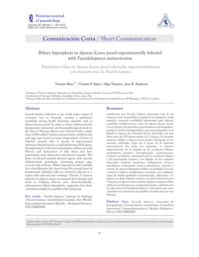
P Peruvian journal J P of parasitology Volumen 20- Número 1 - Año 2012 ISSN 2311-4533 (Electronic version) Comunicación Corta / Short Communication Biliary hyperplasia in alpacas (Lama pacos) experimentally infected with Fasciolahepatica metacercariae Hiperplasia biliar en alpacas (Lama pacos) infectadas experimentalmente con metacercarias de Fasciola hepatica 1, 2* 2 3 3 Vicente Maco , Vicente P. Maco , Olga Timoteo , Jose R. Espinoza 1.Institute of Tropical Medicine Alexander von Humboldt, Cayetano Heredia University (UPCH), Lima, Peru. 2.Laboratory of Anatomic Pathology, Guadalupe Clinic, Lima, Peru. 3.Institute Molecular Biotechnology Unit, Laboratories for Research and Development,Schoolof Science & Philosophy, UPCH, Lima,Peru. Abstract Resumen Fasciola hepatica infection is one of the major causes of economic loss in livestock, causing a significant morbidity among South American camelids such as alpacas (Lama pacos). In order to better understand the host-parasite interaction and histopathological insult in the liver, 6 Huacaya alpacas were infected with a single dose of 200 viable F. hepatica metacercariae. Adult worms and eggs were found in tissue preparations of livers of infected animals after 6 month of experimental infection. Hepatic lesions as wall thickening of bile ducts, disorganization of the liver parenchyma, stellate scar with fibrosis and destruction of bile ducts and liver parenchyma were observed in all infected animals. The livers of infected animals showed regions with chronic inflammation, granuloma containing parasite eggs, necrosis and cirrhosis. Biliary hyperplasia with multiple foci of proliferative bile ducts formed by several layers of transformed epithelial cells was observed adjacent to a region with extensive liver damage. Chronic F. hepatica infection in alpacas causes an extensive liver damage and leads to bridging fibrosis and, characteristically, adenomatous biliary hyperplasia, suggesting that these camelids are highly susceptible to this trematode. Lainfección por Fasciola hepatica representa una de las mayores causas de perdidas económicas en el ganado a nivel mundial, causando morbilidad importante entre algunos camélidos sudamericanos como las alpacas (Lama pacos). Con el objetivo de entender major la interacción hospederoparásito y el daño histopatológico que causa el parásito en el hígado, 6 alpacas tipo Huacaya fueron infectadas con una dosis única de 200 metacercarias de F. hepatica. Se visualizó parásitos adultos y huevos en los tejidos del hígado de los animales infectados luego de 6 meses de la infection experimental. En todos los animales, se observó engrosamiento de las paredes de los conductos biliares, parénquima hepático desorganizado, cicatrizaciones estelares con fibrosis y destrucción de los conductos biliares y del parénquima hepático. Los hígados de los animales infectados también mostraron inflamacion crónica, granulomas conteniendo huevos parasitarios, necrosis y cirrosis. Se observó hiperplasia biliar con múltiples focos de conductos biliares proliferativos formados por múltiples capas de células epiteliales transformadas, adyacentes a la región con daño hepático extenso. La infeccióncrónica por F. hepatica en alpacas causa un daño hepático extensor y lleva a fibrosis portal extensa y,característicamente, a laformación de adenomas de hiperplasia biliar, lo cual sugiere que estos camélidos son altamente susceptibles a la infección por este distoma. Key words: Fasciola hepatica/ anatomy & histology |Fasciola hepática /metabolism|Camelids, New World| Immunohistochemistry|Models, Biological|(Source: DECS BIREME) Palabras clave: Fasciola hepatica /anatomía & histopatología| Fasciola hepatica /metabolismo |Camélidos Americanos| Inmunohistoquímica| Modelos Biológicos| (Fuente: DECS BIREME) Citation: Maco V, Maco VP, Timoteo O, Espinoza JR Biliary hyperplasia in alpacas (Lama pacos) experimentally infected with Fasciola hepatica metacercariae. Peru j parasitol. 2012;20(1): e25-e29 25 Comunicación Corta Maco et al. Biliary hyperplasia in alpacas infected with Fasciola hepatica Peruvian journal of parasitology Volumen 20- Número 1 - Año 2012 ISSN 2311-4533 (Electronic version) F asciola hepatica (F. hepatica) infection is one of the major causes of economic loss in livestock worldwide, with a great impact in the economy of endemic areas in Peru.2A wide range of domestic species is susceptible to the liver fluke infection, as alpacas (Lama pacos), in which the infection causes anemia, eosinophilia, weight loss, emaciation and mortality.3 Despite the importance of this infection by its negative impact in fiber productivity and quality, little is known of the liver pathology caused by F. hepatica in South American camelids, and of the parasite factors that produce the pathogenesis of the disease. aged 18 to 24 months) from the department of Junin (Central Highlands of Peru) were infected with a single dose of 200 metarcariae of F. hepatica administered orally, after a 30-days adaption period. The metacercariae were obtained from lymnaeid snails infected with laboratory-raised F. hepatica miracidia. Prior to inoculation, fluke eggs were not detected in stool samples by the Rapid Sedimentation Technique (RST) by Lumbreras,8 and enzyme-linked immunosorbent assays (ELISA) using Fas1, Fas2 and excretory/secretory products were negative.5 All of the infected alpacas and 1 uninfected control underwent necropsy 6 months after the first single dose.The study protocol was approved by the Institutional Ethics Committee for Animals. Tissue samples from livers were fixed in formaldehyde, included in paraffin and processed by hematoxylin-eosin (H&E) and Masson's trichrome (MT) stains, the latter used to differentiate between collagen fibers and smooth muscle from tumors. Von Kossa, a stain intended for use in the evaluation of calcium deposits, was not performed since standard H&E is considered highly sensitive for mineralization in anatomopathology. The following variables were recorded: portal fibrosis, bile duct hyperplasia, cirrhosis, presence of eggs, granulomas, and the severity of lesions were graded as mild (fibrosis: enlarged fibrotic portal tract; hyperplasia: 25%-50% epithelial height), moderate (fibrosis: periportal fibrosis or portal to portal fibrosis; hyperplasia: 50%-75% epithelial height), and severe (fibrosis: bridging fibrosis with architectural distorsion; hyperplasia >75% epithelial height). A differential susceptibility of the host to the fluke is a feature of the disease, being cattle able to mount a strong immunological response that immunoprotects to subsequent infections, and sheep develop little or absent immunoprotective response to F. hepatica infection .4 It is not known whether alpacas become resistant to F. hepatica after a primary infection. However, field studies suggest that animals acquiredmultiple infections in endemic areas and they are re-infected after treatment with 5 flukicide. Alpacas produce antibody response to liver fluke infection, which peaks 8 weeks after primary infection, but this response seems unable to confer immunoprotection to reinfection and does not prevent the liver damage observed in infected animals.6 U n t i l n o w, n o d e s c r i p t i o n o f t h e histopathological insults caused by the fluke infection was reported in alpacas, as it is well described in sheep, cows and goats. 7In alpacas, the liver penetration appears to start 4 weeks after infection suggested by the elevation of hepatic enzymes observed in infected animals and bile ducts resident mature flukes shed eggs 8 weeks after the infection. 6 F. hepatica infection of alpacas was confirmed by the visualization of eggs (Fig. 1) and adult worms (not shown) in the liver tissue sections. Gross anatomy of the livers of infected animals showed hepatomegaly, fibrosis and calcified bile ducts. Matureflukes were recovered from the livers atnecropsy. The intensity of infection was recorded according to the parasite load. The parasite load averaged 41± 4 (18%-25%) from an infective dose of 200 metacercaria e per animal. In order to gain insight into the histopathological changes of the fluke in the liver and biliary track of this South American camelids, 6 Huacaya alpacas (4 males, 2 females, Peruv. j. parasitol 2012; 20 (1) Acceso gratuito en línea a texto completo. 26 Comunicación Corta Maco et al. Biliary hyperplasia in alpacas infected with Fasciola hepatica Peruvian journal of parasitology Volumen 20- Número 1 - Año 2012 ISSN 2311-4533 (Electronic version) E E (b) (a) Figure 1. (a) Multiple characteristics Fasciola hepatica eggs shells (E) in bile ducts (H&E 40X). (b) Magnification of parasitic elements in situ (H&E 200X). Scale bars: 100nm. H & Esections of infected liver tissue showed extensive tracks of fibrosis (Fig. 2b) as a response to the liver damage caused by the fluke.The infection produced an extensive liver damage that ranges from inflammatory response to cirrhosis. The liver of infected animals showed patches of acute and chronic inflammation to the liver fluke and to the eggs. Granuloma containing parasite ova was a common finding in H&E sections of the liver with chronic infection (Fig. 3a). Inflammatory response recruited lymphocytes, macrophages and plasma cells. We did not detected eosinophils in the inflammatory foci, in spite that peripheral blood eosinophilia in blood was observed in infected alpacas. A peripheral ring of fibroblasts with multinucleated giant cells, lymphocytes and plasma cells surrounded granuloma containing F.hepatica ova (Fig. 3b). Multinucleated cell and lymphocytes were abundant in mineralized granuloma and in areas with cirrhosis. Mild to severe grades of bile duct hyperplasia with biliary adenoma formation (Fig. 4). C FB N FB (b) (a) Figure 2. (a) Necrosis (N) and calcification (C) in the liver of an alpaca experimentally infected with Fasciola hepatica metacercariae. Portal triad shows sclerosed bile duct and calcification (H&E 40X). (b) Different degrees of fibrosis, with nodular to bridging fibrosis (FB), and cirrhosis (MT 40X). Scale bars: 100nm Peruv. j. parasitol 2012; 20 (1) Acceso gratuito en línea a texto completo. 27 Comunicación Corta Maco et al. Biliary hyperplasia in alpacas infected with Fasciola hepatica Peruvian journal of parasitology Volumen 20- Número 1 - Año 2012 ISSN 2311-4533 (Electronic version) The presence of multiple non-encapsulated biliary adenomes in infected animals is a feature of this infection not described in other species.7 The neoplastic tissue consists of biliary epithelial proliferation with no evidence of nuclear or cellular atypia or the presence of mitotic figures. Multiple foci of proliferative bile ducts formed by several layers of transformed epithelial cells were observed djacent to a region with extensive liver damage (Figs. 4a and 4b). A severe form of neoplasia that produced the atrophy of the liver right lobe was caused by the hyperplasia of biliary epithelia was previously described in F. hepatica infected alpaca. 9A similar finding, hyperplasia of the gall bladder was described in a man with a chronic infection.10The development of biliary hyperplasia associated to F. hepatica infection is probably not an uncommon event andit is suggestive of the high susceptibility of the alpacas and other hoststo the liver fluke infection. G E (a) I (b) Figure 3. (a) Portal triads and many hystiocytes forming epithelioid granulomas (G) (H&E 200X). (b) Portal leukocytic infiltration (I) and dilatation of bile ducts with presence of an egg (E) in the lumen (H&E 200 X).Scale bars: 100nm. (b) Different degrees of fibrosis, with nodular to bridging fibrosis (FB), and cirrhosis (MT 40X). Scale bars: 100nm In conclusion, alpacas are a highly susceptible species to F. hepatica assuggested by the extensive liver damage with a distortion of the liverarchitecture that even leads to tissue transformation with characteristics of biliary adenomes in experimentally infected animals. (b) (a) Figure 4. (a) Prominent adenomatous biliary hyperplasia in alpacas (H&E 40X) with magnification b) (H&E 200X). Scale bars: 500nm (4a) and 100nm (4b). (H&E 200 X).Scale bars: 100nm. (b) Different degrees of fibrosis, with nodular to bridging fibrosis (FB), and cirrhosis (MT 40X). Scale bars: 100nm Peruv. j. parasitol 2012; 20 (1) Acceso gratuito en línea a texto completo. 28 Comunicación Corta Maco et al. Biliary hyperplasia in alpacas infected with Fasciola hepatica Peruvian journal of parasitology Volumen 20- Número 1 - Año 2012 ISSN 2311-4533 (Electronic version) Author’s contribution: VM, OT, and JRE Acknowlegdments. This work was partially designed the study. VM and VPM performed all histopathological stains and analyzed the paraffin-embedded liver tissues. funded by a grant B/2856-1 from the International Foundation for Science an INCAGRO CONTRACT 007-2003 to JRE. OT received support from the Consortium of Francophones Universities of Belgium (CIUF) and RED SAREC. E. Chavarry for providing the Fas2 antiserum. Conflict of Interest: The authors declare none. References 1. Spithill TW, Smooker PM, Sexton JL, Bozas E, Morrison CA, Creaney J, Parsons JC. Development of Vaccines Against Fasciola hepatica. In: Dalton JP, editor. Fasciolosis. Cambridge, UK: CABI Publishing; 1999. p. 377-401. 6.Timoteo O, Maco VJr., Maco V, Neyra V, Yi PJ, Leguia G, Espinoza JR. Characterization of the humoral immune response in alpacas (Lama pacos) experimentally infected with Fasciola hepatica against cysteine proteinases Fas1 and Fas2 and h i st o p a t h o l o g i c a l f i n d i n g s . Vet I m m u n o l Immunopathol. 2005; 106: 77-86. 2. Espinoza JR, Terashima A, Herrera-Velit P, Marcos LA. [Human and animal fascioliasis in Peru: impact in the economy of endemic zones]. Rev Peru Med Exp Salud Publica. 2010; 27: 604-612. 7.Behm CA, Sangster NC. Pathology, Pathophysiology and Clinical Aspects. In: Dalton JP, editor. Fasciolosis. Cambridge, UK: CABI Publishing; 1999. p. 377-401. 3. Leguia G, Casas E. Distomatosis hepatica. In: Diaz FS, editor. Parasitic diseases and parasitological atlas of South American camelids. First ed. Lima, Peru: Editorial de Mar; 1999. p. 40-63. 8.Lumbreras H, Cantella R, Burga R, 1962. Acerca de un procedimiento de sedimentación rápida para investigar huevos de Fasciola hepatica en las heces, su evaluación y uso en el campo. Rev Méd Peruana. 1962; 31: 167-74. 4.Mulcahy G, Joyce P, Dalton JP. Immunology of Fasciola hepatica Infection. In: Dalton JP, editor. Fasciolosis. Cambridge, UK: CABI Publishing; 1999. p. 377-401. 9.Hamir AN, Smith BB. Severe biliary hyperplasia associated with liver fluke infection in an adult alpaca. Vet Pathol. 2002; 39: 592-594. 5.Neyra V, Chavarry E, Espinoza JR. Cysteine proteinases Fas1 and Fas2 are diagnostic markers for Fasciola hepatica infection in alpacas (Lama pacos). Vet Parasitol. 2002; 105: 21-32. 10.Osman M, Lausten SB, El-Sefi T, Boghdadi I, Rashed MY, Jensen SL. Biliary parasites. Dig Surg. 1998; 15: 287-296. Correspondence: Vicente Maco. Current address: Honorio Delgado 430, Urb. Ingenieria, San Martin de Porres, Lima, Peru. Tel.: +1 202 247 0505. E-mail address: vicente_maco@hotmail.com Peruv. j. parasitol 2012; 20 (1) Acceso gratuito en línea a texto completo. 29

