Verrucous Carcinoma of the Face - Actas Dermo
Anuncio
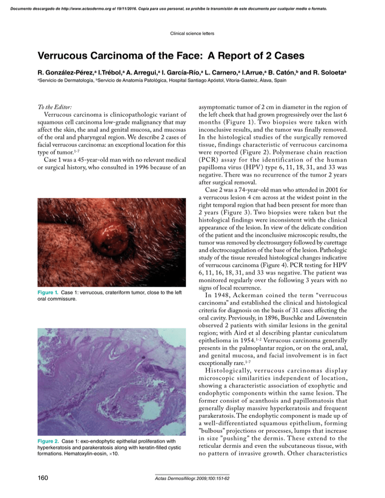
Documento descargado de http://www.actasdermo.org el 19/11/2016. Copia para uso personal, se prohíbe la transmisión de este documento por cualquier medio o formato. Clinical science letters Verrucous Carcinoma of the Face: A Report of 2 Cases R. González-Pérez,a I.Trébol,a A. Arregui,a I. García-Río,a L. Carnero,a I.Arrue,a B. Catón,b and R. Soloetaa Servicio de Dermatología, bServicio de Anatomía Patológica, Hospital Santiago Apóstol, Vitoria-Gasteiz, Álava, Spain a To the Editor: Verrucous carcinoma is clinicopathologic variant of squamous cell carcinoma low-grade malignancy that may affect the skin, the anal and genital mucosa, and mucosas of the oral and pharyngeal region. We describe 2 cases of facial verrucous carcinoma: an exceptional location for this type of tumor.1-7 Case 1 was a 45-year-old man with no relevant medical or surgical history, who consulted in 1996 because of an Figure 1. Case 1: verrucous, crateriform tumor, close to the left oral commissure. Figure 2. Case 1: exo-endophytic epithelial proliferation with hyperkeratosis and parakeratosis along with keratin-filled cystic formations. Hematoxylin-eosin, ×10. 160 asymptomatic tumor of 2 cm in diameter in the region of the left cheek that had grown progressively over the last 6 months (Figure 1). Two biopsies were taken with inconclusive results, and the tumor was finally removed. In the histological studies of the surgically removed tissue, findings characteristic of verrucous carcinoma were reported (Figure 2). Polymerase chain reaction (PCR) assay for the identification of the human papilloma virus (HPV ) type 6, 11, 18, 31, and 33 was negative. There was no recurrence of the tumor 2 years after surgical removal. Case 2 was a 74-year-old man who attended in 2001 for a verrucous lesion 4 cm across at the widest point in the right temporal region that had been present for more than 2 years (Figure 3). Two biopsies were taken but the histological findings were inconsistent with the clinical appearance of the lesion. In view of the delicate condition of the patient and the inconclusive microscopic results, the tumor was removed by electrosurgery followed by curettage and electrocoagulation of the base of the lesion. Pathologic study of the tissue revealed histological changes indicative of verrucous carcinoma (Figure 4). PCR testing for HPV 6, 11, 16, 18, 31, and 33 was negative. The patient was monitored regularly over the following 3 years with no signs of local recurrence. In 1948, Ackerman coined the term “verrucous carcinoma” and established the clinical and histological criteria for diagnosis on the basis of 31 cases affecting the oral cavity. Previously, in 1896, Buschke and Löwenstein observed 2 patients with similar lesions in the genital region; with Aird et al describing plantar cuniculatum epithelioma in 1954.1-2 Verrucous carcinoma generally presents in the palmoplantar region, or on the oral, anal, and genital mucosa, and facial involvement is in fact exceptionally rare.1-7 Histologic al l y, verr ucous c arcinomas display microscopic similarities independent of location, showing a characteristic association of exophytic and endophytic components within the same lesion. The former consist of acanthosis and papillomatosis that generally display massive hyperkeratosis and frequent parakeratosis. The endophytic component is made up of a well-differentiated squamous epithelium, forming ”bulbous” projections or processes, lumps that increase in size ”pushing” the dermis. These extend to the reticular dermis and even the subcutaneous tissue, with no pattern of invasive growth. Other characteristics Actas Dermosifiliogr. 2009;100:151-62 Documento descargado de http://www.actasdermo.org el 19/11/2016. Copia para uso personal, se prohíbe la transmisión de este documento por cualquier medio o formato. Clinical science letters include the formation of keratin cysts and the presence of a variable inflammatory infiltrate. 1,2 Clinical and histological differential diagnosis should mainly include other variants of squamous carcinoma, common warts, keratoacanthomas, and reactive epidermal hyperplasias. Less commonly, they can be confused with certain benign adnexal tumors, giant seborrheic keratoses, verrucous melanomas, verruciform xanthomas, and even with iododermas and bromodermas. 2 W here ver diagnostic doubt exists, the complete removal of the tumor is required in order to reach a correct diagnosis as biopsies alone cannot show the ‘exo-endophytic’ growth characteristic of verrucous carcinoma. In the 2 cases presented, biopsies were taken from the lesions on several occasions giving contradictory results. Diagnosis of 1 of the cases was further complicated by the keratoacanthomatous clinical appearance of the lesion. In both cases complete excision of the tumor was required for definitive diagnosis. The pathogenesis of the disease has classically involved chemical factors like chewing tobacco—in the case of Ackerman tumor—and various others including chronic inflammation. More recently, the identification of HPV by in situ hybridization or PCR in some of these tumors— mainly on the mucosa—has prompted consideration of the virus as another pathogenic agent. Both low risk (HPV 6 and 11) and high risk (HPV 16 and 18) genotypes have been implicated here. In fact, HPV 6 and 11 are characteristically related to Buschke-Löwenstein carcinoma, while other forms (HPV 16, 18, and 33) have also been demonstrated in verrucous carcinomas in other locations. 1,2,8,9 In our 2 patients PCR did not identify HPV. This is in agreement with the findings of many other authors who confirm that it is rare to find these viruses in verrucous carcinomas in zones outside those traditionally affected.2,8,10 In summary, we have described 2 cases of verrucous carcinoma of the facial area—a ver y rare location considering there are only 5 cases previously published. 3-7 PCR for the identification of HPV was not undertaken in any of those 5 cases. PCR did not reveal presence of HPV in either of our patients, a fact that suggests the appearance of verrucous carcinomas in such light-exposed locations is the outcome of different pathogenic factors—mainly ultraviolet radiation—as is the case in other cutaneous squamous cell carcinomas. Figure 3. Case 2: clinical appearance of the verrucous carcinoma. Figure 4. Case 2: epithelial tumor of mixed exophytic-verrucous and endophytic growth, consisting of atypical squamous cells. Hematoxylin-eosin, ×10. Correspondence: Ricardo González Pérez Servicio de Dermatología Hospital Santiago Apóstol C/ Olaguibel 29 01004 Vitoria-Gasteiz, Álava, Spain RICARDO.GONZALEZPEREZ@osakidetza.net Conflicts of Interest The authors declare no conflicts of interest. References Acknowledgments We thank Drs Luis Requena and Evaristo Sánchez Yus for their valuable collaboration in the review of the histological samples in Case 1. 1. McKee PH, Calonje E, Granter RC. Pathology of the Skin with clinical correlations. 3rd ed. London: Elsevier; 2005. p. 1218-20. 2. Schwartz RA. Verrucous carcinoma of the skin and mucosa. J Am Acad Dermatol. 1995;32:1-21. Actas Dermosifiliogr. 2009;100:151-62 161 Documento descargado de http://www.actasdermo.org el 19/11/2016. Copia para uso personal, se prohíbe la transmisión de este documento por cualquier medio o formato. Clinical science letters 3. Gallo A, Fiorella ML, Simonelli M, Rocca CD, Vincentiis M. Carcinoma cuniculatum: verrucous carcinoma of the skin of the face. Otolaryngology-Head and Neck Surgery. 2005;133:1-2. 4. Nguyen KQ, McMarlin SL. Verrucous carcinoma of the face. Arch Dermatol. 1984;120:383-5. 5. Takematsu H, Watanabe M, Matsunaga J, Ueno H, Tagami H. Verrucous carcinoma of the face with a massive neutrophil infiltrate. Analysis of leukocyte chemotactic activity in the tumour extract. Clin Exp Dermatol. 1994;19: 26-30. 6. Shimizu A, Tamura A, Ishikawa O. Invasive squamous cell carcinoma arising from verrucous carcinoma. Recognition of 162 7. 8. 9. 10. verrucous carcinoma of skin as an in situ carcinoma. Eur J Dermatol. 2006;16:439-42. W ilkinson JD, Martin M, Black MM. Carcinoma cuniculatum: A clinicopathologic study of 21 cases. Arch Dermatol.1980;116:1390. Pattee SF, Bordeaux J, Mahalingam M, Nitzan YB, Maloney ME. Verrucous carcinoma of the scalp. J Am Acad Dermatol.2007;56:506-7. Corbalán-Vélez R, Ruiz-Maciá JA, Brufau C, Carapeto FJ. Carcinoma espinocelular cutáneo y papilomavirus (VPH). Actas Dermosifiliogr. 2007;98:583-93. Murao K, Kubo Y, Fukumoto D, Matsumoto K, Arase S. Verrucous carcinoma of the scalp associated with human papillomavirus type 33. Dermatol Surg. 2005;31:1363-5. Actas Dermosifiliogr. 2009;100:151-62
