Management of Basal Cell Carcinoma with Perineural Invasion
Anuncio
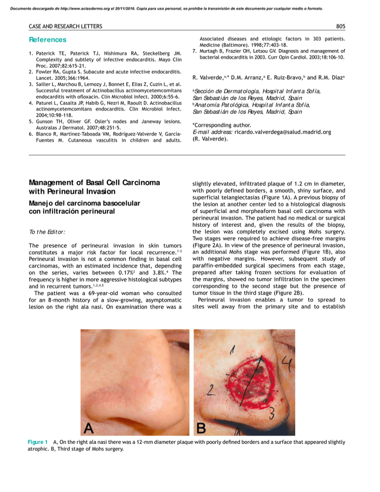
Documento descargado de http://www.actasdermo.org el 20/11/2016. Copia para uso personal, se prohíbe la transmisión de este documento por cualquier medio o formato. CASE AND RESEARCH LETTERS References 1. Paterick TE, Paterick TJ, Nishimura RA, Steckelberg JM. Complexity and subtlety of infective endocarditis. Mayo Clin Proc. 2007;82:615-21. 2. Fowler RA, Gupta S. Subacute and acute infective endocarditis. Lancet. 2005;366:1964. 3. Sailler L, Marchou B, Lemozy J, Bonnet E, Elias Z, Cuzin L, et al. Successful treatment of Actinobacillus actinomycetemcomitans endocarditis with oloxacin. Clin Microbiol Infect. 2000;6:55-6. 4. Paturel L, Casalta JP, Habib G, Nezri M, Raoult D. Actinobacillus actinomycetemcomitans endocarditis. Clin Microbiol Infect. 2004;10:98-118. 5. Gunson TH, Oliver GF. Osler’s nodes and Janeway lesions. Australas J Dermatol. 2007;48:251-5. 6. Blanco R, Martínez-Taboada VM, Rodríguez-Valverde V, GarcíaFuentes M. Cutaneous vasculitis in children and adults. Management of Basal Cell Carcinoma with Perineural Invasion Manejo del carcinoma basocelular con infiltración perineural To t he Edit or: The presence of perineural invasion in skin tumors constitutes a major risk factor for local recurrence.1-3 Perineural invasion is not a common finding in basal cell carcinomas, with an estimated incidence that, depending on the series, varies between 0.17%2 and 3.8%.4 The frequency is higher in more aggressive histological subtypes and in recurrent tumors.1,2,4,5 The patient was a 69-year-old woman who consulted for an 8-month history of a slow-growing, asymptomatic lesion on the right ala nasi. On examination there was a 805 Associated diseases and etiologic factors in 303 patients. Medicine (Baltimore). 1998;77:403-18. 7. Murtagh B, Frazier OH, Letsou GV. Diagnosis and management of bacterial endocarditis in 2003. Curr Opin Cardiol. 2003;18:106-10. R. Valverde,a,* D.M. Arranz,a E. Ruiz-Bravo,b and R.M. Díaza Sección de Dermat ología, Hospit al Inf ant a Sof ía, San Sebast ián de los Reyes, Madrid, Spain b Anat omía Pat ológica, Hospit al Inf ant a Sof ía, San Sebast ián de los Reyes, Madrid, Spain a *Corresponding author. E-mail address: ricardo.valverdega@salud.madrid.org (R. Valverde). slightly elevated, infiltrated plaque of 1.2 cm in diameter, with poorly defined borders, a smooth, shiny surface, and superficial telangiectasias (Figure 1A). A previous biopsy of the lesion at another center led to a histological diagnosis of superficial and morpheaform basal cell carcinoma with perineural invasion. The patient had no medical or surgical history of interest and, given the results of the biopsy, the lesion was completely excised using Mohs surgery. Two stages were required to achieve disease-free margins (Figure 2A). In view of the presence of perineural invasion, an additional Mohs stage was performed (Figure 1B), also with negative margins. However, subsequent study of paraffin-embedded surgical specimens from each stage, prepared after taking frozen sections for evaluation of the margins, showed no tumor infiltration in the specimen corresponding to the second stage but the presence of tumor tissue in the third stage (Figure 2B). Perineural invasion enables a tumor to spread to sites well away from the primary site and to establish Figure 1 A, On the right ala nasi there was a 12-mm diameter plaque with poorly deined borders and a surface that appeared slightly atrophic. B, Third stage of Mohs surgery. Documento descargado de http://www.actasdermo.org el 20/11/2016. Copia para uso personal, se prohíbe la transmisión de este documento por cualquier medio o formato. 806 CASE AND RESEARCH LETTERS Figure 2 A, Supericial basal cell carcinoma in the irst Mohs stage (Hematoxylin-eosin, original magniication ×100). B, Residual tumor deposits (arrows). Dermal iniltrate of basaloid cells interspersed between the hair follicles (Hematoxylin-eosin, original magniication ×250). an independent lesion with clinical, histological, and pathogenic characteristics that distinguish it both from local spread and from vascular or lymphatic invasion.6 Histologically, perineural dissemination is characterized by a patchy pattern with areas of normal tissue between foci of neoplastic infiltration.4,6,7 This characteristic pattern, which some authors consider to be merely an artifact of processing and fixing the specimen, explains the persistence of tumor in apparently healthy tissue despite correct examination of all the margins.8 Due to the high risk of local recurrence, basal cell carcinomas with perineural invasion require specific management. The majority of authors agree on the use of Mohs surgery as the treatment of choice for this type of t umor 2-4,7-10; however, the use of other therapeutic options, such as adjuvant radiotherapy2,4,7,9 or performing an additional Mohs stage after obtaining negative margins,4,7,9 continues to be a subject of debate. Unfortunately there are no randomized studies that have compared the different therapeutic options. We consider that the case presented here supports the use of Mohs surgery with an additional stage for the treatment of basal cell carcinoma with perineural invasion. This will ensure the greatest likelihood of complete resection of the tumor and will thus reduce the risk of recurrence. 2. Leibovitch I, Huilgol SC, Selva D, Richards S, Paver R. Basal cell carcinoma treated with Mohs surgery in Australia III. Perineural invasion. J Am Acad Dermatol. 2005;53:458-63. 3. Ríos-Buceta L, Picoto A. Cirugía de Mohs. Actas Dermosiiliogr. 2003;94:503-23. 4. Ratner D, Lowe L, Johnson TM, Fader DJ. Perineural spread of basal cell carcinomas treated with Mohs micrographic surgery. Cancer. 2000;88:1605-13. 5. Brown CI, Perry AE. Incidence of perineural invasion in histologically aggressive types of basal cell carcinoma. Am J Dermatopathol. 2000;22:123-5. 6. Liebig C, Ayala G, Wilks JA, Berger DH, Albo D. Perineural invasion in cancer: a review of the literature. Cancer. 2009;115:3379-91. 7. Feasel AM, Brown TJ, Bogle MA, Tschen JA, Nelson BR. Perineural invasion of cutaneous malignancies. Dermatol Surg. 2001;27:531-42. 8. Matorin PA, Wagner RF. Mohs micrographic surgery: technical dificulties posed by perineural invasion. Int J Dermatol. 1992;31:83-6. 9. Geist DE, Garcia-Moliner M, Fitzek MM, Cho H, Rogers GS. Perineural invasion of cutaneous squamous cell carcinoma and basal cell carcinoma: raising awareness and optimizing management. Dermatol Surg. 2008;34:1642-51. 10. Telfer NR, Colver GB, Morton CA. Guidelines for the management of basal cell carcinoma. Br J Dermatol. 2008;159:35-48. References Servicio de Dermat ología, Inst it ut o Valenciano de Oncología, Valencia, Spain 1. Batra RS, Kelley LC. Predictors of extensive subclinical spread in nonmelanoma skin cancer treated with Mohs micrographic surgery. Arch Dermatol. 2002;138:1043-51. F. Messeguer,* E. Nagore-Enguídanos, C. Requena, and C. Guillén-Barona *Corresponding author. E-mail address: francescmb@comv.es (F. Messeguer).
