A Rare Termination of Left Common Facial Vein into Left
Anuncio
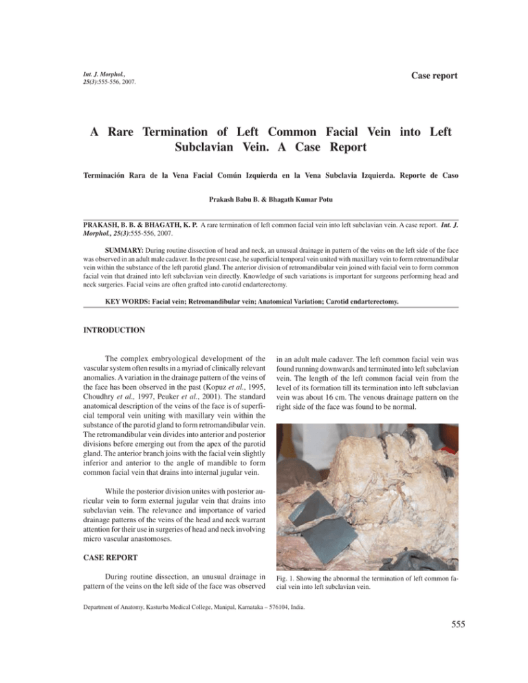
Int. J. Morphol., 25(3):555-556, 2007. Case report A Rare Termination of Left Common Facial Vein into Left Subclavian Vein. A Case Report Terminación Rara de la Vena Facial Común Izquierda en la Vena Subclavia Izquierda. Reporte de Caso Prakash Babu B. & Bhagath Kumar Potu PRAKASH, B. B. & BHAGATH, K. P. A rare termination of left common facial vein into left subclavian vein. A case report. Int. J. Morphol., 25(3):555-556, 2007. SUMMARY: During routine dissection of head and neck, an unusual drainage in pattern of the veins on the left side of the face was observed in an adult male cadaver. In the present case, he superficial temporal vein united with maxillary vein to form retromandibular vein within the substance of the left parotid gland. The anterior division of retromandibular vein joined with facial vein to form common facial vein that drained into left subclavian vein directly. Knowledge of such variations is important for surgeons performing head and neck surgeries. Facial veins are often grafted into carotid endarterectomy. KEY WORDS: Facial vein; Retromandibular vein; Anatomical Variation; Carotid endarterectomy. INTRODUCTION The complex embryological development of the vascular system often results in a myriad of clinically relevant anomalies. A variation in the drainage pattern of the veins of the face has been observed in the past (Kopuz et al., 1995, Choudhry et al., 1997, Peuker et al., 2001). The standard anatomical description of the veins of the face is of superficial temporal vein uniting with maxillary vein within the substance of the parotid gland to form retromandibular vein. The retromandibular vein divides into anterior and posterior divisions before emerging out from the apex of the parotid gland. The anterior branch joins with the facial vein slightly inferior and anterior to the angle of mandible to form common facial vein that drains into internal jugular vein. in an adult male cadaver. The left common facial vein was found running downwards and terminated into left subclavian vein. The length of the left common facial vein from the level of its formation till its termination into left subclavian vein was about 16 cm. The venous drainage pattern on the right side of the face was found to be normal. While the posterior division unites with posterior auricular vein to form external jugular vein that drains into subclavian vein. The relevance and importance of varied drainage patterns of the veins of the head and neck warrant attention for their use in surgeries of head and neck involving micro vascular anastomoses. CASE REPORT During routine dissection, an unusual drainage in pattern of the veins on the left side of the face was observed Fig. 1. Showing the abnormal the termination of left common facial vein into left subclavian vein. Department of Anatomy, Kasturba Medical College, Manipal, Karnataka – 576104, India. 555 PRAKASH, B. B. & BHAGATH, K. P. A rare termination of left common facial vein into left subclavian vein. A case report. Int. J. Morphol., 25(3):555-556, 2007. DISCUSSION Deviations in the venous system from the normal pattern are quite common. Like most superficial veins, the external veins of the head and neck are subject to variations in their morphology, size and termination. Variations in the course and termination of the facial veins are not uncommon (Bergman, 1998). Kopuz, described an unusual course of the facial vein which joined the retromandibular vein at a higher level in the parotid gland on the right side of the face Choudhry found that the termination of the facial vein in the external jugular vein occurs in 5% of the individuals. To the best of our knowledge the termination of the common facial vein in the subclavian vein has so far not been documented in the available literature. Knowledge of the varying venous patterns in the region of the face is important for the surgeons in order to avoid any intra-operative trial and error procedures which might lead to unnecessary bleeding (Nagase et al., 1997). These facial veins may be used as patches for carotid endarterectomy (Sabharwal & Mukherjeed, 1998) and for oral reconstruction. Remembering the varying venous patterns in the head and face region is important to preoperatively evaluate the course and branching patterns of the respective vessels, by means of Doppler Ultrasonography. The embryological basis of the present variation is not known since it is the first case reported so far in the available literature. PRAKASH, B. B. & BHAGATH, K. P. Terminación rara de la vena facial común izquierda en la vena subclavia izquierda. Reporte de caso. Int. J. Morphol., 25(3):555-556, 2007. RESUMEN: Durante una disección de rutina de cabeza y cuello, fue observado en un cadáver aduto masculino un inusual drenaje de los patrones venosos del lado izquierda de la cara. La vena temporal superficial se unía con la vena maxilar formando la vena retromandibular dentro del parénquima de la glándula parótida izquierda. La división anterior de la vena retromandibular se unió a la vena facial formando la vena facial común la cual drenó directamente en la vena subclavia izquierda. El conocimiento de estas variaciones es importante para los cirujanos de cabeza y cuello. A menudo, las venas faciales se injertan en la endarterectomía carotídea. PALABRAS CLAVE: Vena facial; Vena retromandibular; Variación anatómica; Endarterectomía carotídea. REFERENCES Bergman, R. A.; Thompson, S. A.; Afifi, A. K. & Saadeh, F. A. Compendium of human anatomic variation: catalog, atlas and world literature. Baltimore:Urban & Schwarzenberg, 1988. Sabharwal, P. & Mukherjeed, D. Autogenous commomn facial vein or external jugular vein patch for carotid endarterectomy. Cardiovascular Surgery, 6:594-597, 1998. Choudhry, R.; Tuli, A. & Choudry, S. Facial vein terminating in the external jugular vein. An embryological interpretation. Surg. Radiol. Anat.,19:73-7, 1997. Correspondence to: Dr. Prakash Babu. B, Associate Professor of Anatomy, Department of Anatomy, Centre for Basic Sciences Kasturba Medical College, Madhav Nagar, Manipal-576104 Karnataka, INDIA. Kopuz, C.; Yavuz, S.; Cumhur, M.; Tftik, S. & IIgi, S. An unusual coursing of the facial vein. Kaibogaku Zasshi, 70: 20-2, 1995. Nagase, T.; Kobayashi, S.; Sekiya, S. & Ohmori, K. Anatomic evaluation of the facial artery and vein using color Doppler ultrasonography. Annals of Plastic Surgery, 39:64-7, 1997. Peuker, E. T.; Fischer, G. & Filler, T. J. Correspondence – facial vein terminating in the superficial temporal vein: a case report. J. Anat., 198:509-10, 2001. 556 Phone: 91 -0820-2922327 Fax : 91-0820-2570061 E-mail:billakantibabu@yahoo.co.in Received: 05-04-2007 Accepted: 15-05-2007 Int. J. Morphol., 25(3):557-560, 2007. Estudio Estereológico del Hígado de Cobayo (Cavia porcellus) Stereological Study of the Guinea-pig Liver (Cavia porcellus) * Carlos Rosas C.; **Bélgica Vásquez & *Mariano del Sol ROSAS, C. C.; VÁSQUEZ, B. & DEL SOL, M. Estudio estereológico del hígado de cobayo (Cavia porcellus). Int. J. Morphol., 25(3):557-560, 2007. RESUMEN: El hígado es un órgano ampliamente investigado por la infinidad de funciones que posee. Por otra parte, la Estereología permite obtener datos cuantitativos de la estructura en estudio. Tener acceso a este tipo de datos de los hepatocitos y sinusoides en condiciones fisiológicas normales, es necesario para determinar como varía el número de estas células en condiciones patológicas. El objetivo principal de esta investigación fue describir la morfología del hígado de cobayo a través de la Estereología, sentando las bases para futuras investigaciones morfofuncionales. Se utilizaron 5 machos adultos de la especie Cavia porcellus, a los cuales se les extrajo el hígado y se determinó su volumen aplicando el método de Scherle. Posteriormente, se obtuvieron 5 trozos de forma aleatoria, los que fueron fijados en solución de Bouin acuoso. De estas muestras se obtuvieron cortes de 3µm, que fueron teñidos con hematoxilina-eosina. Para el estudio estereológico, se usó el sistema M42. Fueron determinados: las densidades de número, volumen y superficie y número total de hepatocitos y sinusoides. Se consideró un índice de significancia menor o igual a 0,05. El NV de hepatocitos fue de 9,20 x 10 5 /mm3, VV de 70,04% y el SV de 254,95 mm2/mm3. En el caso de los sinusoides el NV fue de 1,94 x 10 5 /mm3, VV de 24,86% y el SV de 47,01 mm2/mm3. El número total de hepatocitos fue de 2,8 x 10⁷ y sinusoides 0,61 x 10⁷. Al comparar los resultados obtenidos en Cavia porcellus y los obtenidos en otras especies, se observa una variación cuantitativa en la morfología hepática. Con estos resultados es posible establecer bases para investigaciones experimentales. PALABRAS CLAVE: Hígado; Cobayo; Cavia porcellus; Estereología. INTRODUCCIÓN El hígado, ocupa un importante papel en la homeostasis del organismo a través de una innumerable cantidad de funciones que realiza. Del punto de vista clínico, es importante detectar alteraciones morfológicas de este órgano para corroborar la existencia de alguna patología. Así, el conocimiento de esta estructura, en especial sus aspectos celulares cuantitativos, resulta fundamental para comprender si el órgano está normal o alterado. Es aquí donde la Estereología juega un rol importante, ya que permite realizar una reconstrucción tridimensional y una estimación cuantitativa de estructuras a nivel microscópico. En relación al número de hepatocitos, se han encontrado diferencias notables entre especies. Rohr et al. (1976) determinaron un 79,3% de hepatocitos en el hígado de seres humanos; en primates Leontopithecus, Burity et al. (2004) obtuvieron 90,1- 94,9%, por otra parte; Weibel et al. (1969) informaron un 83,1%, en el hígado de ratas Wistar; y Blouin et al. (1977) un 77,8%, en el parénquima del hígado de ratas Sprague-Dawley. Por otra parte, Águila et al. (2003) en estudios experimentales en ratas, alimentadas con una dieta alta en lípidos, determinaron que el número de hepatocitos se encontraba entre 56 y 62%. Si bien es cierto, se han realizado numerosos estudios del hígado de varias especies, en los cuales se ha analizado la acción de diversos agentes, hay pocos en la especie Cavia porcellus. Respecto a los sinusoides hepáticos, algunos estudios morfométricos han sido realizados en diversas especies. Ragonha et al. (2006) determinaron que en el hígado de ratones, un 18,3% correspondía a sinusoides. * ** Facultad de Medicina, Universidad de La Frontera, Temuco, Chile. Universidad Autónoma de Chile, Temuco, Chile 557 ROSAS, C. C.; VÁSQUEZ, B. & DEL SOL, M. Estudio estereológica del hígado de cobayo (Cavia porcellus). Int. J. Morphol., 25(3):557-560, 2007. El objetivo del estudio, fue determinar algunos aspectos cuantitativos del hígado de cobayo (Cavia porcellus), a través de la estereología. Así, con la generación de estos nuevo conocimientos nos será posible sentar las bases para futuras investigaciones morfofuncionales. MATERIAL Y MÉTODO Utilizamos 5 cobayos machos adultos (Cavia porcellus), con un peso promedio de 711g (DE ± 212), sanos y mantenidos ad libitum en el Bioterio de la Universidad de La Frontera, Temuco, Chile. Los animales fueron sacrificados por traumatismo encéfalo craneano. A través de una laparotomía, fue retirado el hígado y se determinó su volumen por el método de Scherle (1970). Obtuvimos 5 trozos, aleatoriamente, siguiendo las reglas del Orientator (Mattfeldt et al.,1990). Las secciones fueron fijadas en Bouin acuoso, deshidratadas e incluidas en parafina para obtener cortes histológicos de un grosor de 3 µm (t = 3 µm), que fueron teñidos con HE. Se analizaron 5 campos por lámina, 125 campos en total. Las láminas fueron observadas en un microscopio óptico Olympus® modelo CX31, con cámara marca Moticam® modelo 480. Las imágenes fueron proyectadas en un monitor de pantalla plana marca Sony®. Para realizar la estereología se usó el test multipropósito M42. Los parámetros estereológicos medidos fueron: densidad de volumen (Vv), densidad de superficie (Sv), densidad numérica (Nv) y número total (n) de hepatocitos y sinusoides hepáticos. Se calcularon, el promedio, la desviación estándar , el error estándar, el coeficiente de variación y el coeficiente de error. Se consideró un índice de significancia menor o igual a 0,05. RESULTADOS El volumen promedio de los hígados de los 5 animales fue de 31,81 mm3. (DE ± 13,20). El número total de hepatocitos fue de 2,8 x 10⁷ y de sinusoides 0,61 x 10⁷ . Los parámetros estereológicos estudiados en hepatocitos y sinusoides se presentan en la Tabla I y las Figs. 1, 2 y 3. DISCUSIÓN Estudios estereológicos en el hígado de Cavia porcellus no se encuentran en la literatura consultada, siendo necesario comparar nuestros resultados con las de otras especies investigadas. El Nv de hepatocitos en cobayo es inferior al obtenido en ratas por Weibel et al. (1969), Carthew et al. (1998) y Martins et al. (2005) y superior a lo informado por Águila et al. (2003), Moreti et al. (2005); Silva et al. (2006), Marcos et al y Ragonha et al., en ratas. En el caso de los primates Leontopithecus, Burity et al. obtuvieron un Nv de hepatocitos muy superior al del cobayo. Con respecto al Vv, los valores en cobayo no son similares a los estudios realizados en otras especies. El 70,04% de hepatocitos determinado en Cavia porcellus, es significativamente inferior a lo informado por Weibel et al. (83,1% de hepatocitos) en ratas Wistar y Blouin et al. (77,8%) en el parénquima del hígado de ratas Sprague-Dawley. Por otra parte, Rohr et al. determinaron un 79,3% de hepatocitos en seres humanos; sin embargo, éstos últimos agregan que el parénquima corresponde al 92,4% del volumen del hígado. Por tanto, el Vv efectivo para el hígado correspondería a un 71,89% de hepatocitos, resultado bastante similar al obtenido en nuestro estudio. Águila et al. determinaron un VV entre 56 y 62%, en ratas que presentaban una dieta alta en lípidos, porcentaje un poco más bajo que el de nuestros resultados. Por otra parte, cuando se compara el VV de sinusoides, los resultados son bastante similares. En el caso de los primates Leontopithecus, Burity et al. obtuvieron en todas las especies estudiadas, un Vv de hepatocitos mucho mayor (90,1- 94,9%) a los obtenidos en Cavia porcellus. Estos autores hacen mención al estudio de Carrière et al. (1980) en hígado de Papio papio, quienes determinaron un Vv de 84,9% de hepatocitos, resultado superior a los obtenidos en los hepatocitos de hígado de cobayo. Moreti et al. encontraron que el hígado de feto de rata Wistar presentaba hepatocitos con un 30,72% de citoplasma y un 9,47% de núcleo, resultados muy inferiores, si se comparan con el VV obtenido en los hepatocitos de cobayo. Ragonha et al. determinaron en el hígado de ratones, un 76,17% de hepatocitos y Tabla I. Parámetros estereológicos del hígado de cobayo (Cavia porcellus). Densidades promedios De número de hepatocitos 9 ,20 x 105 célul as/m m3 70,04% De volumen de hepatocitos De superficie de hepatocitos 254,95 mm2/ mm3 De número de sinusoides hepáticos 1,94 x 10⁵ sinusoides/mm3 De volumen de sinusoides hepáticos 24,86% 2 3 De superficie de sinusoides hepáticos 47,01 mm / mm 558 DE CV (%) EE CE (%) ±2,4 x 10⁵ ±8,91 31,39 0,43 x 10⁵ 26,03 12,73 12,31 1,07 x 10⁵ 3,98 14,01 0,19 x 10⁵ 11,6 5,68 5,50 ±3,6 2 3 1 0,63 mm /mm 21,97 20,01 22,60 1,61 4,74 9,81 6,46 10,09 ROSAS, C. C.; VÁSQUEZ, B. & DEL SOL, M. Estudio estereológica del hígado de cobayo (Cavia porcellus). Int. J. Morphol., 25(3):557-560, 2007. un 18,3% de sinusoides, resultados similares a los nuestros. En cambio, si comparamos los resultados con Neves et al. (2006), obtenidos en ratones Swiss Webster, estos últimos presentan VV de hepatocitos más elevado. Fig. 1. Número total de hepatocitos y sinusoides hepáticos en 5 hígados de cobayo (Cavia porcellus). Se pudo comprobar que existen fluctuaciones en el número total de hepatocitos y sinusoides por cobayo estudiado, debido a que el volumen total del hígado de cada ejemplar era diferente. Sin embargo, el Nv en cada uno de ellos fue muy similar, siendo este último dato mucho más trascendental a la hora de hacer comparaciones con otras especies. Las diferencias obtenidas en otras investigaciones podrían deberse, tanto a la diversidad de técnicas que existen para determinar los parámetros morfológicos y estereológicos, como también a los efectos producidos por los estudios experimentales, edad y condiciones medioambientales de los animales. Con los resultados obtenidos en esta investigación se han sentado las bases para futuros estudios experimentales que se puedan realizar en el hígado del cobayo Cavia porcellus. Fig. 2. Densidad de volumen de hepatocitos y sinusoides hepáticos en 5 hígados de cobayo (Cavia porcellus). Fig. 3. Densidad de superficie de hepatocitos y sinusoides hepáticos en 5 hígados de cobayo (Cavia porcellus). ROSAS, C. C.; VÁSQUEZ, B. & DEL SOL, M. Stereological study of the guinea-pig liver (Cavia porcellus). Int. J. Morphol., 25(3):557560, 2007. SUMMARY: The liver is a widely researched organ for having an infinity of functions. On the other hand, Stereology allows the access of quantitaive data of the study´s structure. Having access to this tpe of date of hepatocitos and sinusoids Under normal physiological conditions, it is necessary to determine how the number of these cells varies in pathological conditions. The principal objective of this investigation was to describe the morphology of the liver of cobayo, through Stereology, setting the bases for future morphofuncional investigations. Five adult males of the Cavia porcellus species were used, of which the liver was extracted and its volume determined, applying the Scherle method. Subsequently five pieces of aleatoria form were obtained which were set in an aqueous Bouin 559 ROSAS, C. C.; VÁSQUEZ, B. & DEL SOL, M. Estudio estereológica del hígado de cobayo (Cavia porcellus). Int. J. Morphol., 25(3):557-560, 2007. solution. Of these samples 3µm cuts were obtained, that were dyed with hematoxilina-eosina. For the stereological study the M42 system was used. The following were determined: the number densities, volume and surface and total number of hepatocitos and sinusoides. An index of lesser significance or equal to 0,05. The NV of hepatocitos was 9.20 x 10⁵ /mm3, VV de 70.04% and SV of 254.95 mm2/mm3. In the case of the sinusoides N V was 1.94 x 10⁵ /mm3, VV of 24.86% and SV de 47.01 mm2/mm3. The total number of hepatocites was of 2.8 x 10⁷ and sinusoides 0.61 x 10⁷. While comparing the results obtained in Cavia porcellus and those obtained in other species, a quantitative variation in the hepatic morphology is observed. With these results it is possible to establish the bases for experimental research . KEY WORDS: Liver; Guinea pig; Cavia porcellus; Stereology. REFERENCIAS BIBLIOGRÁFICAS Águila, M. B.; Pinheiro, A. R.; Parente, L. B. & Mandarimde-Lacerda, C. A. Dietary effect of different high-fat diet on rat liver stereology. Liver International, 23: 363-70, 2003. Blouin, A.; Bolender, R. P. & Weibel, E. R. Distribution of organelles and membranes between hepatocytes and nonhepatocytes in rat liver parenchyma – a stereological study. J. Cell. Biol., 72:441-55, 1977. Burity, C. H. F.; Pissinatti, A. & Mandarim-de-Lacerda, C. A. Stereology of the Liver in three Species of Leontopithecus (Lesson, 1840) Callitrichidae – Primates. Anat. Histol. Embryol., 33:183-7, 2004. Carthew, P.; Edwards, R. E. & Nolan, B. M. The quantitative distinction of hyperplasia from hypertrophy in hepatomegaly induced in the rat liver by phenobarbital. Toxicol. Sci., 44:46-51, 1998. Moreti, D. L. C.; Lopes, R. A.; Vinha, D.; Sala, M. A.; Semprini, M. & Friedrichi, C. Efectos del albendazol en el hígado de feto de rata. Estudios morfológico y morfométrico. Int. J. Morphol., 23(2):111-120, 2005. Neves, R. H.; Alencar, A. C.; Aguila, M. B.; Mandarim-deLacerda, C. A.; Machado-Silva, J. R. & Gomes, D. C. Hepatic stereology of schistosomiasis mansoni infectedmice fed a high-fat diet. Mem. Inst. Oswaldo Cruz, 101(1):253-60, 2006. Ragonha, L. H. O.; Abrahao, A. A. C.; Sala, M. A.; Lopes, R. A.; Ribeiro, R. D.; Prado JR., J. C.; Albuquerque, S. & Zucoloto, S. Estudio morfométrico del hígado de ratón en la enfermedad de Chagas experimental. Int. J. Morphol., 24(3):383-90, 2006. Rohr, H. P.; Luthy, J.; Gudat, F.; Oberholzer, M.; Gysin, C. & Bianchi, L. Stereology of liver biopsies from healthy volunteers. Virchows Arch. A Path. Anat. And Histol., 371:251-63, 1976. Scherle W. A simple method for volumetry of organs in quantitative stereology. Mikroskopie, 26:57-60,1970. Silva, R. F.; Lopes, R. A.; Sala, M. A.; Vinha, D.; Regalo, S. C. H.; Souza, A. M. & Gregorio, Z. M. O. Action of trivalent chromium on rat liver structure. Histometric and haematological studies. Int. J. Morphol., 24(2):197203, 2006. Weibel, E. R.; Staubli W.; Gnagi H. R. & Hess F. A. Correlated morphometric and biochemical studies on the liver cell – I. Morphometric model, stereologic methods, and normal morphometric data for rat liver. J. Cell Biol., 42:68-91, 1969. Marcos, R.; Monteiro, R. A. F. & Rocha, E. Design-based stereological estimation of hepatocyte number, by combing the smooth optical fractionator and immunocytochemistry with anti-carcinoembryonic antigen polyclonal antibodies. Liver International, 26: 116-24, 2006. Dirección para correspondencia: Prof. Dr. Mariano del Sol C. Facultad de Medicina Universidad de La Frontera Casilla 54-D Temuco - CHILE Martins, A. T.; Azoubel, R.; Lopes, R. A.; Di Matteo, M. A. S. & Arruda, J. G. F. Effect of sodium cyclamate on the rat fetal liver: A karyometric and stereological study. Int. J. Morphol., 23(3):221-6, 2005. Email: mdelsol@ufro.cl Mattfeldt, T.; Mall, G.; Gharehbaghi, H. & Moller, P. Estimation of surface area and length with the Orientator. J. Microsc., 159 (3):301-17, 1990. 560 Recibido: 22-03-2007 Aceptado: 27-07-2007
