18F FDG PET/CT in a child with gliomatosis cerebri
Anuncio

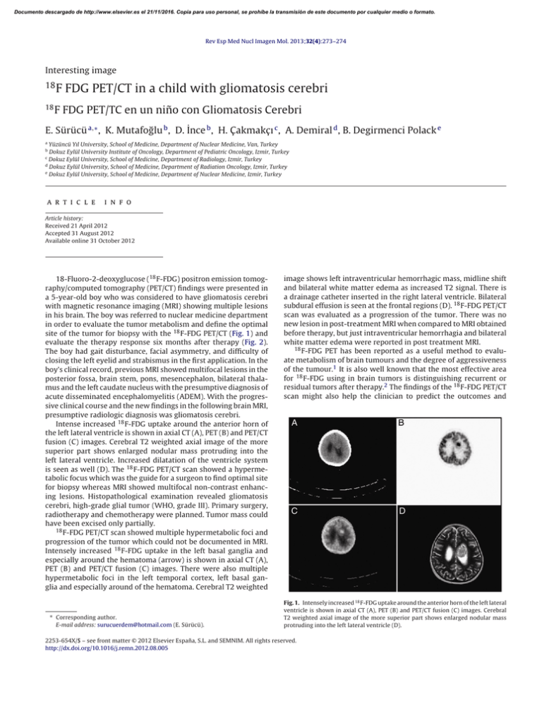
Documento descargado de http://www.elsevier.es el 21/11/2016. Copia para uso personal, se prohíbe la transmisión de este documento por cualquier medio o formato. Rev Esp Med Nucl Imagen Mol. 2013;32(4):273–274 Interesting image 18 18 F FDG PET/CT in a child with gliomatosis cerebri F FDG PET/TC en un niño con Gliomatosis Cerebri E. Sürücü a,∗ , K. Mutafoğlu b , D. İnce b , H. Çakmakçı c , A. Demiral d , B. Degirmenci Polack e a Yüzüncü Yıl University, School of Medicine, Department of Nuclear Medicine, Van, Turkey Dokuz Eylül University Institute of Oncology, Department of Pediatric Oncology, Izmir, Turkey c Dokuz Eylül University, School of Medicine, Department of Radiology, Izmir, Turkey d Dokuz Eylül University, School of Medicine, Department of Radiation Oncology, Izmir, Turkey e Dokuz Eylül University, School of Medicine, Department of Nuclear Medicine, Izmir, Turkey b a r t i c l e i n f o Article history: Received 21 April 2012 Accepted 31 August 2012 Available online 31 October 2012 18-Fluoro-2-deoxyglucose (18 F-FDG) positron emission tomography/computed tomography (PET/CT) findings were presented in a 5-year-old boy who was considered to have gliomatosis cerebri with magnetic resonance imaging (MRI) showing multiple lesions in his brain. The boy was referred to nuclear medicine department in order to evaluate the tumor metabolism and define the optimal site of the tumor for biopsy with the 18 F-FDG PET/CT (Fig. 1) and evaluate the therapy response six months after therapy (Fig. 2). The boy had gait disturbance, facial asymmetry, and difficulty of closing the left eyelid and strabismus in the first application. In the boy’s clinical record, previous MRI showed multifocal lesions in the posterior fossa, brain stem, pons, mesencephalon, bilateral thalamus and the left caudate nucleus with the presumptive diagnosis of acute disseminated encephalomyelitis (ADEM). With the progressive clinical course and the new findings in the following brain MRI, presumptive radiologic diagnosis was gliomatosis cerebri. Intense increased 18 F-FDG uptake around the anterior horn of the left lateral ventricle is shown in axial CT (A), PET (B) and PET/CT fusion (C) images. Cerebral T2 weighted axial image of the more superior part shows enlarged nodular mass protruding into the left lateral ventricle. Increased dilatation of the ventricle system is seen as well (D). The 18 F-FDG PET/CT scan showed a hypermetabolic focus which was the guide for a surgeon to find optimal site for biopsy whereas MRI showed multifocal non-contrast enhancing lesions. Histopathological examination revealed gliomatosis cerebri, high-grade glial tumor (WHO, grade III). Primary surgery, radiotherapy and chemotherapy were planned. Tumor mass could have been excised only partially. 18 F-FDG PET/CT scan showed multiple hypermetabolic foci and progression of the tumor which could not be documented in MRI. Intensely increased 18 F-FDG uptake in the left basal ganglia and especially around the hematoma (arrow) is shown in axial CT (A), PET (B) and PET/CT fusion (C) images. There were also multiple hypermetabolic foci in the left temporal cortex, left basal ganglia and especially around of the hematoma. Cerebral T2 weighted ∗ Corresponding author. E-mail address: surucuerdem@hotmail.com (E. Sürücü). image shows left intraventricular hemorrhagic mass, midline shift and bilateral white matter edema as increased T2 signal. There is a drainage catheter inserted in the right lateral ventricle. Bilateral subdural effusion is seen at the frontal regions (D). 18 F-FDG PET/CT scan was evaluated as a progression of the tumor. There was no new lesion in post-treatment MRI when compared to MRI obtained before therapy, but just intraventricular hemorrhagia and bilateral white matter edema were reported in post treatment MRI. 18 F-FDG PET has been reported as a useful method to evaluate metabolism of brain tumours and the degree of aggressiveness of the tumour.1 It is also well known that the most effective area for 18 F-FDG using in brain tumors is distinguishing recurrent or residual tumors after therapy.2 The findings of the 18 F-FDG PET/CT scan might also help the clinician to predict the outcomes and Fig. 1. Intensely increased 18 F-FDG uptake around the anterior horn of the left lateral ventricle is shown in axial CT (A), PET (B) and PET/CT fusion (C) images. Cerebral T2 weighted axial image of the more superior part shows enlarged nodular mass protruding into the left lateral ventricle (D). 2253-654X/$ – see front matter © 2012 Elsevier España, S.L. and SEMNIM. All rights reserved. http://dx.doi.org/10.1016/j.remn.2012.08.005 Documento descargado de http://www.elsevier.es el 21/11/2016. Copia para uso personal, se prohíbe la transmisión de este documento por cualquier medio o formato. 274 E. Sürücü et al. / Rev Esp Med Nucl Imagen Mol. 2013;32(4):273–274 prognosis of the patient with the primary glial brain tumors.3 In this particular case, 18 F-FDG PET/CT scan added very valuable diagnostic information to the MRI in terms of guidance of biopsy site and documenting progressive disease after the therapy. Unfortunately, the child died with progression of disease at the 10th month of oncologic diagnosis. References 1. Di Chiro G, DeLaPaz RL, Brooks RA, Sokoloff L, Kornblith PL, Smith BH, et al. Glucose utilization of cerebral gliomas measured by [18F] fluorodeoxyglucose and positron emission tomography. Neurology. 1982;32:1323–9. 2. Ho LT, Wassef H, Henderson R, Seto J. F-18 FDG PET-CT imaging in recurrent cerebral gliosarcoma. Clin Nucl Med. 2009;34:153–4. 3. Alavi JB, Alavi A, Chawluk J, Kushner M, Powe J, Hickey W, et al. Positron emission tomography in patients with glioma. A predictor of prognosis. Cancer. 1988;62:1074–8. Fig. 2. Intensely increased 18 F-FDG uptake in the left basal ganglia and especially around of the hematoma (arrow) is shown in axial CT (A), PET (B) and PET/CT fusion (C) images. Cerebral T2 weighted image shows left intraventricular hemorrhagic mass, midline shift and bilateral white matter edema as increased T2 signal (D).


