ESCRS survey reveals risk factors for post
Anuncio
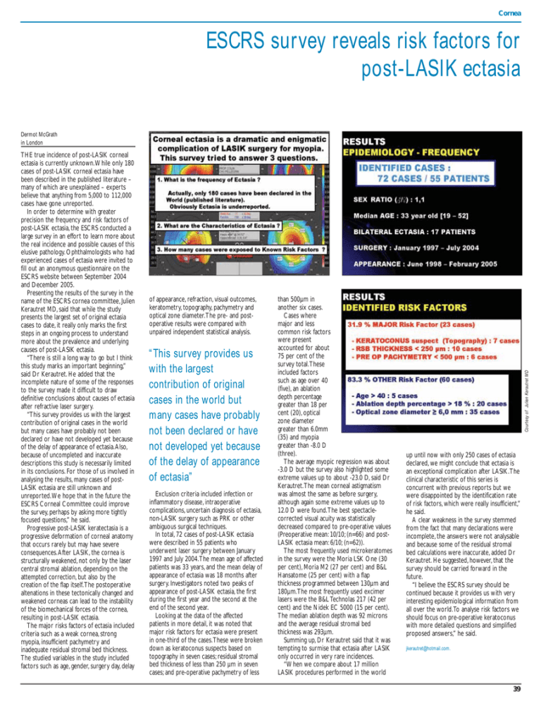
Cornea ESCRS survey reveals risk factors for post-LASIK ectasia THE true incidence of post-LASIK corneal ectasia is currently unknown.While only 180 cases of post-LASIK corneal ectasia have been described in the published literature – many of which are unexplained – experts believe that anything from 5,000 to 112,000 cases have gone unreported. In order to determine with greater precision the frequency and risk factors of post-LASIK ectasia, the ESCRS conducted a large survey in an effort to learn more about the real incidence and possible causes of this elusive pathology. Ophthalmologists who had experienced cases of ectasia were invited to fill out an anonymous questionnaire on the ESCRS website between September 2004 and December 2005. Presenting the results of the survey in the name of the ESCRS cornea committee, Julien Kerautret MD, said that while the study presents the largest set of original ectasia cases to date, it really only marks the first steps in an ongoing process to understand more about the prevalence and underlying causes of post-LASIK ectasia. “There is still a long way to go but I think this study marks an important beginning,” said Dr Kerautret. He added that the incomplete nature of some of the responses to the survey made it difficult to draw definitive conclusions about causes of ectasia after refractive laser surgery. “This survey provides us with the largest contribution of original cases in the world but many cases have probably not been declared or have not developed yet because of the delay of appearance of ectasia.Also, because of uncompleted and inaccurate descriptions this study is necessarily limited in its conclusions. For those of us involved in analysing the results, many cases of postLASIK ectasia are still unknown and unreported.We hope that in the future the ESCRS Corneal Committee could improve the survey, perhaps by asking more tightly focused questions,” he said. Progressive post-LASIK keratectasia is a progressive deformation of corneal anatomy that occurs rarely but may have severe consequences.After LASIK, the cornea is structurally weakened, not only by the laser central stromal ablation, depending on the attempted correction, but also by the creation of the flap itself.The postoperative altenations in these tectonically changed and weakened corneas can lead to the instability of the biomechanical forces of the cornea, resulting in post-LASIK ectasia. The major risks factors of ectasia included criteria such as a weak cornea, strong myopia, insufficient pachymetry and inadequate residual stromal bed thickness. The studied variables in the study included factors such as age, gender, surgery day, delay of appearance, refraction, visual outcomes, keratometry, topography, pachymetry and optical zone diameter.The pre- and postoperative results were compared with unpaired independent statistical analysis. “This survey provides us with the largest contribution of original cases in the world but many cases have probably not been declared or have not developed yet because of the delay of appearance of ectasia” Exclusion criteria included infection or inflammatory disease, intraoperative complications, uncertain diagnosis of ectasia, non-LASIK surgery such as PRK or other ambiguous surgical techniques. In total, 72 cases of post-LASIK ectasia were described in 55 patients who underwent laser surgery between January 1997 and July 2004.The mean age of affected patients was 33 years, and the mean delay of appearance of ectasia was 18 months after surgery. Investigators noted two peaks of appearance of post-LASIK ectasia, the first during the first year and the second at the end of the second year. Looking at the data of the affected patients in more detail, it was noted that major risk factors for ectasia were present in one-third of the cases.These were broken down as keratoconus suspects based on topography in seven cases; residual stromal bed thickness of less than 250 µm in seven cases; and pre-operative pachymetry of less than 500µm in another six cases. Cases where major and less common risk factors were present accounted for about 75 per cent of the survey total.These included factors such as age over 40 (five), an ablation depth percentage greater than 18 per cent (20), optical zone diameter greater than 6.0mm (35) and myopia greater than -8.0 D (three). The average myopic regression was about -3.0 D but the survey also highlighted some extreme values up to about -23.0 D, said Dr Kerautret.The mean corneal astigmatism was almost the same as before surgery, although again some extreme values up to 12.0 D were found.The best spectaclecorrected visual acuity was statistically decreased compared to pre-operative values (Preoperative mean: 10/10; (n=66) and postLASIK ectasia mean: 6/10; (n=62)). The most frequently used microkeratomes in the survey were the Moria LSK One (30 per cent), Moria M2 (27 per cent) and B&L Hansatome (25 per cent) with a flap thickness programmed between 130µm and 180µm.The most frequently used excimer lasers were the B&L Technolas 217 (42 per cent) and the Nidek EC 5000 (15 per cent). The median ablation depth was 92 microns and the average residual stromal bed thickness was 293µm. Summing up, Dr Kerautret said that it was tempting to surmise that ectasia after LASIK only occurred in very rare incidences. “When we compare about 17 million LASIK procedures performed in the world Courtesy of Julien Kerautret MD Dermot McGrath in London up until now with only 250 cases of ectasia declared, we might conclude that ectasia is an exceptional complication after LASIK.The clinical characteristic of this series is concurrent with previous reports but we were disappointed by the identification rate of risk factors, which were really insufficient,” he said. A clear weakness in the survey stemmed from the fact that many declarations were incomplete, the answers were not analysable and because some of the residual stromal bed calculations were inaccurate, added Dr Kerautret. He suggested, however, that the survey should be carried forward in the future. “I believe the ESCRS survey should be continued because it provides us with very interesting epidemiological information from all over the world.To analyse risk factors we should focus on pre-operative keratoconus with more detailed questions and simplified proposed answers,” he said. jkerautret@hotmail.com. 39
