Enlargement of the thigh and osteoblastic lesion in a young man
Anuncio

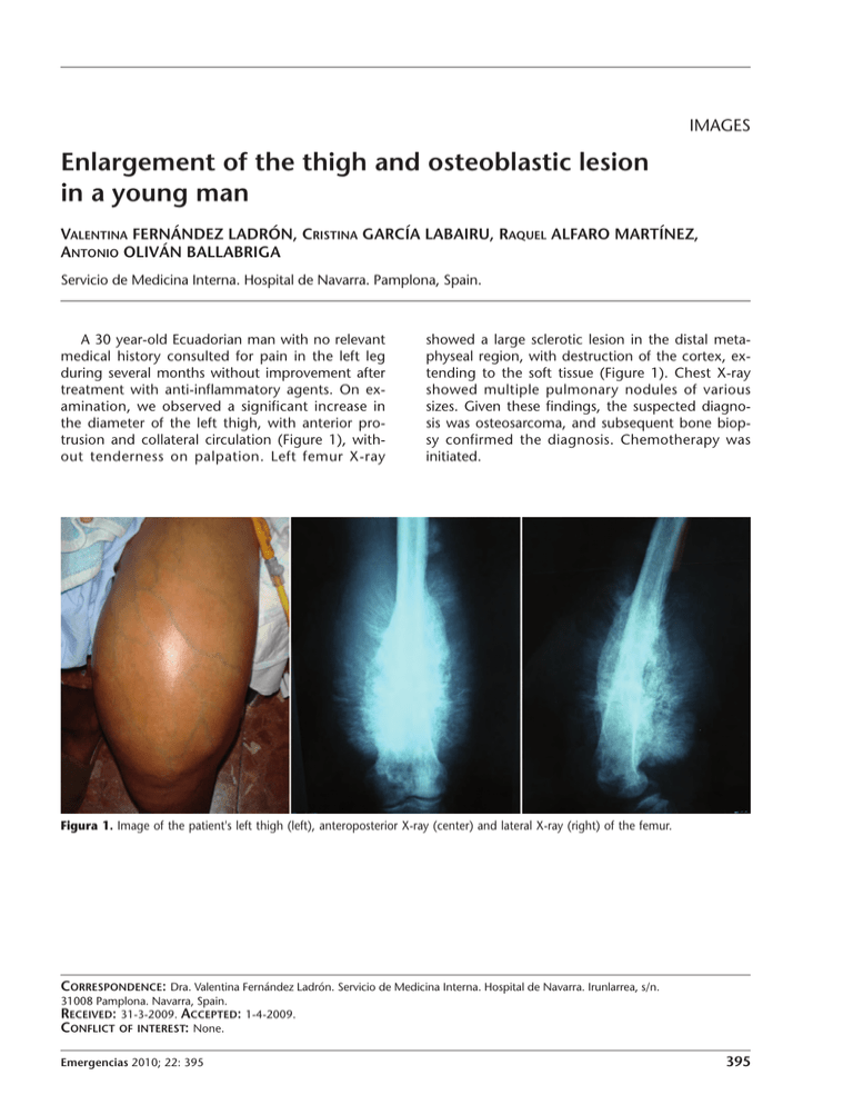
IMAGES Enlargement of the thigh and osteoblastic lesion in a young man VALENTINA FERNÁNDEZ LADRÓN, CRISTINA GARCÍA LABAIRU, RAQUEL ALFARO MARTÍNEZ, ANTONIO OLIVÁN BALLABRIGA Servicio de Medicina Interna. Hospital de Navarra. Pamplona, Spain. A 30 year-old Ecuadorian man with no relevant medical history consulted for pain in the left leg during several months without improvement after treatment with anti-inflammatory agents. On examination, we observed a significant increase in the diameter of the left thigh, with anterior protrusion and collateral circulation (Figure 1), without tenderness on palpation. Left femur X-ray showed a large sclerotic lesion in the distal metaphyseal region, with destruction of the cortex, extending to the soft tissue (Figure 1). Chest X-ray showed multiple pulmonary nodules of various sizes. Given these findings, the suspected diagnosis was osteosarcoma, and subsequent bone biopsy confirmed the diagnosis. Chemotherapy was initiated. Figura 1. Image of the patient's left thigh (left), anteroposterior X-ray (center) and lateral X-ray (right) of the femur. CORRESPONDENCE: Dra. Valentina Fernández Ladrón. Servicio de Medicina Interna. Hospital de Navarra. Irunlarrea, s/n. 31008 Pamplona. Navarra, Spain. E-mail: fernandezvalentina@hotmail.com RECEIVED: 31-3-2009. ACCEPTED: 1-4-2009. CONFLICT OF INTEREST: None. Emergencias 2010; 22: 395 395


