Sickle Cell Trait Splenic Infarction: Mt. Fuji Case Report
Anuncio
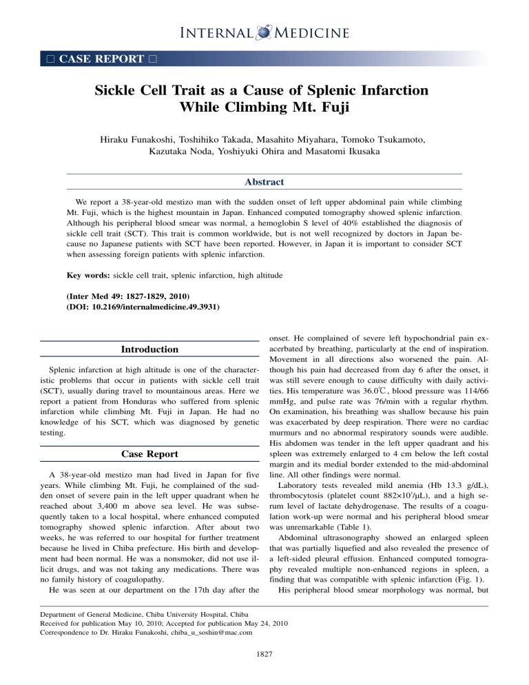
□ CASE REPORT □ Sickle Cell Trait as a Cause of Splenic Infarction While Climbing Mt. Fuji Hiraku Funakoshi, Toshihiko Takada, Masahito Miyahara, Tomoko Tsukamoto, Kazutaka Noda, Yoshiyuki Ohira and Masatomi Ikusaka Abstract We report a 38-year-old mestizo man with the sudden onset of left upper abdominal pain while climbing Mt. Fuji, which is the highest mountain in Japan. Enhanced computed tomography showed splenic infarction. Although his peripheral blood smear was normal, a hemoglobin S level of 40% established the diagnosis of sickle cell trait (SCT). This trait is common worldwide, but is not well recognized by doctors in Japan because no Japanese patients with SCT have been reported. However, in Japan it is important to consider SCT when assessing foreign patients with splenic infarction. Key words: sickle cell trait, splenic infarction, high altitude (Inter Med 49: 1827-1829, 2010) (DOI: 10.2169/internalmedicine.49.3931) Introduction Splenic infarction at high altitude is one of the characteristic problems that occur in patients with sickle cell trait (SCT), usually during travel to mountainous areas. Here we report a patient from Honduras who suffered from splenic infarction while climbing Mt. Fuji in Japan. He had no knowledge of his SCT, which was diagnosed by genetic testing. Case Report A 38-year-old mestizo man had lived in Japan for five years. While climbing Mt. Fuji, he complained of the sudden onset of severe pain in the left upper quadrant when he reached about 3,400 m above sea level. He was subsequently taken to a local hospital, where enhanced computed tomography showed splenic infarction. After about two weeks, he was referred to our hospital for further treatment because he lived in Chiba prefecture. His birth and development had been normal. He was a nonsmoker, did not use illicit drugs, and was not taking any medications. There was no family history of coagulopathy. He was seen at our department on the 17th day after the onset. He complained of severe left hypochondrial pain exacerbated by breathing, particularly at the end of inspiration. Movement in all directions also worsened the pain. Although his pain had decreased from day 6 after the onset, it was still severe enough to cause difficulty with daily activities. His temperature was 36.0℃, blood pressure was 114/66 mmHg, and pulse rate was 76/min with a regular rhythm. On examination, his breathing was shallow because his pain was exacerbated by deep respiration. There were no cardiac murmurs and no abnormal respiratory sounds were audible. His abdomen was tender in the left upper quadrant and his spleen was extremely enlarged to 4 cm below the left costal margin and its medial border extended to the mid-abdominal line. All other findings were normal. Laboratory tests revealed mild anemia (Hb 13.3 g/dL), thrombocytosis (platelet count 882×103/μL), and a high serum level of lactate dehydrogenase. The results of a coagulation work-up were normal and his peripheral blood smear was unremarkable (Table 1). Abdominal ultrasonography showed an enlarged spleen that was partially liquefied and also revealed the presence of a left-sided pleural effusion. Enhanced computed tomography revealed multiple non-enhanced regions in spleen, a finding that was compatible with splenic infarction (Fig. 1). His peripheral blood smear morphology was normal, but Department of General Medicine, Chiba University Hospital, Chiba Received for publication May 10, 2010; Accepted for publication May 24, 2010 Correspondence to Dr. Hiraku Funakoshi, chiba_u_soshin@mac.com 1827 Inter Med 49: 1827-1829, 2010 DOI: 10.2169/internalmedicine.49.3931 Ta bl e1 . La bo r a t o r yPr o f i l e Complete blood count WBC Red blood cells Hb Hematocrit Platelet count Coagulation profile Activated partial thromboplastin time Prothrombin time international ratio Protein C Protein S Lupus anticoagulant Anticardiolipin antibody Plasminogen Chemistry Aspartate Transaminase 28 Alanine Transaminase 43 Lactate Dehydrogenase 561 Alkaline phosphatase 479 Gamma-Glutamyltransferase 139 Total Protein 6.5 Albumin 3.5 27.8 second Urea nitrogen 8 1.05 Creatinine 0.88 115 % Sodium 139 125 % Potassium 4.4 negative Cloride 104 <8 U/mL 146 % 10.3 4.63 13.3 38.9 882 ×103/μL ×106/μL g/dL % ×103/μL U/L U/L U/L U/L U/L g/dL g/dL mg/dL U/L mEq/L mEq/L mEq/L Fi g ur e2 . Re s ul t so fhe mo g l o bi ne l e c t r o pho r e s i s .Thel o we r pa ne ls ho wsno r ma la dul the mo l y s a t ea sac o nt r o l .Theuppe r pa ne li st hi spa t i e nt ’ she mo l y s a t ea ndt hea r r o wi ndi c a t e sa n a bno r ma lba nd. Fi g ur e1 . Enha nc e dc o mput e dt o mo g r a phywa spe r f o r me d o nt hef i f t hda ya f t e rt heo ns e ta tt hel o c a lc l i ni ct ha tt hi spa t i e ntf i r s tv i s i t e d.I ts ho wsa ne nl a r g e ds pl e e n( a r r o w)wi t h he t e r o g e ne o use nha nc e me nta ndal e f t s i de dpl e ur a le f f us i o n. that did not exclude SCT. This is because the red blood cells of persons with SCT only develop a sickle shape under hypoxemic conditions, unlike the red cells of persons with sickle cell anemia. Therefore, we requested hemoglobin electrophoresis, which revealed abnormal hemoglobin (Fig. 2). In addition, isoelectric focusing of hemolysate showed the presence of a slow-moving abnormal hemoglobin, and high performance liquid chromatography revealed that the content of this hemoglobin was 40.5%. Structural analysis by genetic testing revealed substitution of the sixth amino acid in the β-globin chain: CAG (glutamic acid [Glu]) to CTG (valine [Val]). Accordingly, the abnormal hemoglobin was identified as hemoglobin S (HbS) (β6 [A3] Glu→Val), and a diagnosis of sickle cell trait was made based on these findings. We managed this patient with rest, analgesia, and other supportive therapy. Conservative treatment was effective and we finished outpatient follow-up at five months after the onset. Discussion Sickle cell disease is the most common structural hemo- globinopathy, especially occurring in African-Americans. This hemoglobinopathy is a compound heterozygous autosomal codominant trait. Sickle cell disease is caused by a single base substitution in the gene encoding the human βglobin subunit that changes the sixth amino acid from glutamic acid to valine. The resulting abnormal Hb, called HbS, polymerizes when deoxygenated and this leads to stiffening of the erythrocyte membrane that causes the characteristic sickle shape of red cells (1). The prototype disease, sickle cell anemia, occurs in persons homozygous for HbS. Most patients with sickle cell anemia suffer from hemolytic anemia. In addition, microinfarction caused by vascular occlusion is sometimes a fatal complication. The diagnosis is established by detecting elongated and crescent-shaped red blood cells on a peripheral blood smear and abnormalities of the complete blood count. In contrast, patients with SCT are usually asymptomatic and not anemic and their peripheral blood smear is normochromic. However, hypoxemia due to strenuous exercise or high altitude causes their red blood cells to become sickleshaped. Because sickle-shaped red blood cells have less plasticity, thrombosis can lead to organ damage such as splenic infarction or pulmonary infarction. The present patient did not know about his SCT and had never experienced any problems, although he often flew in airplanes and climbed mountains about 2,500 m high. The pressure in the cabin of an airplane is 0.75 atmospheres, which corresponds to 2,400 m above sea level. The red 1828 Inter Med 49: 1827-1829, 2010 DOI: 10.2169/internalmedicine.49.3931 blood cells of persons with SCT usually develop a sickle shape at about 3,500 m above sea level in the absence of heavy exercise. This is why he had no symptoms while flying or climbing other mountains, but splenic infarction occurred when he was about 3,400 m above sea level on Mt. Fuji. Splenic infarction is usually a self-limiting condition. It is often associated with a left-sided pleural effusion, while laboratory tests typically show an increase of LDH and mild anemia. Whether the treatment of splenic infarction in patients with sickle cell disease should be splenectomy or conservative management is still under discussion (2), although many reports have indicated that conservative management can be effective (3, 4). The present patient responded well to conservative therapy and there were no complications. We advised our patient with SCT that he should avoid hypoxemic situations such as heavy exercise or high altitude, use oxygen when flying in an airplane, and receive a vaccination for Streptococcus pneumoniae and Haemophilus influenzae infection (5). The prevalence of SCT is especially high among AfricanAmericans, while it is not seen among Japanese (6). Thus, SCT is not familiar to Japanese doctors, although numerous cases are reported overseas. The present case indicates that in Japan it is necessary to consider the possibility of SCT when examining foreign patients with splenic infarction. Every physician working in or around mountainous areas about 3,000 m above sea level, like Mt. Fuji, should be altered to this issue. Conclusion It is important to consider the possibility of SCT when a foreign patient presents with the acute onset of abdominal pain at a high altitude. We cannot rule out SCT, even when the peripheral blood smear is normal, because only hypoxemia causes the red cells of patients with SCT to become sickle-shaped. Accordingly, when SCT is strongly suspected, hemoglobin electrophoresis should be performed. Acknowledgement We are grateful to Dr. Keiko Harano, Department of Biochemistry, Kawasaki Medical School, Kurashiki, Japan for hemoglobin analysis. References 1. Bunn HF. Pathogenesis and treatment of sickle cell disease. N Engl J Med 337: 762-769, 1997. 2. Owusu-Ofuri S, Hirst C. Splenectomy versus conservative management for acute sequestration crises in people with sickle cell disease. Cochrane Database Syst Rev 4: CD003425, 2002. 3. Sheikha A. Splenic syndrome in patients at high altitude with unrecognized sickle cell trait: splenectomy is often unnecessary. Can J Surg 48: 377-381, 2005. 4. Franklin QJ, Compeggie M. Splenic syndrome in sickle cell trait: four case presentations and a review of the literature. Mil Med 164: 230-233, 1999. 5. Clasta S, Vichinsky EP. Managing sickle cell disease. BMJ 327: 1151, 2003. 6. Shibata Y, Koita I, Kishi T, Kizawa S, Hirooka Y. The family case of sickle cell disease. Igaku Kensa 55: 744-748, 2006 (in Japanese, Abstract in English). Ⓒ 2010 The Japanese Society of Internal Medicine http://www.naika.or.jp/imindex.html 1829
