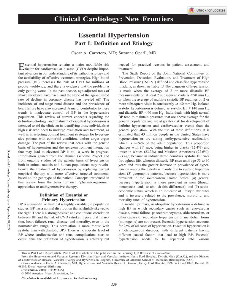
Clinical Cardiology: New Frontiers Essential Hypertension Part I: Definition and Etiology Oscar A. Carretero, MD; Suzanne Oparil, MD E Downloaded from http://ahajournals.org by on September 28, 2024 ssential hypertension remains a major modifiable risk factor for cardiovascular disease (CVD) despite important advances in our understanding of its pathophysiology and the availability of effective treatment strategies. High blood pressure (BP) increases the risk of CVD for millions of people worldwide, and there is evidence that the problem is only getting worse. In the past decade, age-adjusted rates of stroke incidence have risen, and the slope of the age-adjusted rate of decline in coronary disease has leveled off. The incidence of end-stage renal disease and the prevalence of heart failure have also increased. A major contributor to these trends is inadequate control of BP in the hypertensive population. This review of current concepts regarding the definition, etiology, and treatment of essential hypertension is intended to aid the clinician in identifying those individuals at high risk who need to undergo evaluation and treatment, as well as in selecting optimal treatment strategies for hypertensive patients with comorbid conditions and/or target organ damage. The part of the review that deals with the genetic basis of hypertension and the gene/environment interaction that may lead to elevated BP is still a work in progress. Information gained from the Human Genome Project and from ongoing studies of the genetic basis of hypertension both in animal models and human populations may revolutionize the treatment of hypertension by replacing current empirical therapy with more effective, targeted treatments based on the genotype of the patient. Concepts introduced in this review form the basis for such “pharmacogenomic” approaches to antihypertensive therapy. needed for practical reasons in patient assessment and treatment. The Sixth Report of the Joint National Committee on Prevention, Detection, Evaluation, and Treatment of High Blood Pressure (JNC VI) defined and classified hypertension in adults, as shown in Table 1.3 The diagnosis of hypertension is made when the average of 2 or more diastolic BP measurements on at least 2 subsequent visits is ⱖ90 mm Hg or when the average of multiple systolic BP readings on 2 or more subsequent visits is consistently ⱖ140 mm Hg. Isolated systolic hypertension is defined as systolic BP ⱖ140 mm Hg and diastolic BP ⬍90 mm Hg. Individuals with high normal BP tend to maintain pressures that are above average for the general population and are at greater risk for development of definite hypertension and cardiovascular events than the general population. With the use of these definitions, it is estimated that 43 million people in the United States have hypertension or are taking antihypertensive medication, which is ⬇24% of the adult population. This proportion changes with (1) race, being higher in blacks (32.4%) and lower in whites (23.3%) and Mexican Americans (22.6%); (2) age, because in industrialized countries systolic BP rises throughout life, whereas diastolic BP rises until age 55 to 60 years and thus the greater increase in prevalence of hypertension among the elderly is mainly due to systolic hypertension; (3) geographic patterns, because hypertension is more prevalent in the southeastern United States; (4) gender, because hypertension is more prevalent in men (though menopause tends to abolish this difference); and (5) socioeconomic status, which is an indicator of lifestyle attributes and is inversely related to the prevalence, morbidity, and mortality rates of hypertension. Essential, primary, or idiopathic hypertension is defined as high BP in which secondary causes such as renovascular disease, renal failure, pheochromocytoma, aldosteronism, or other causes of secondary hypertension or mendelian forms (monogenic) are not present. Essential hypertension accounts for 95% of all cases of hypertension. Essential hypertension is a heterogeneous disorder, with different patients having different causal factors that lead to high BP. Essential hypertension needs to be separated into various Definition of Essential or Primary Hypertension BP is a quantitative trait that is highly variable1; in population studies, BP has a normal distribution that is slightly skewed to the right. There is a strong positive and continuous correlation between BP and the risk of CVD (stroke, myocardial infarction, heart failure), renal disease, and mortality, even in the normotensive range. This correlation is more robust with systolic than with diastolic BP.2 There is no specific level of BP where cardiovascular and renal complications start to occur; thus the definition of hypertension is arbitrary but This is Part I of a 2-part article. Part II of this article will be published in the February 1, 2000 issue of Circulation. From the Hypertension and Vascular Research Division, Heart and Vascular Institute, Henry Ford Hospital, Detroit, Mich (O.A.C.), and the Division of Cardiovascular Disease, Vascular Biology and Hypertension Program, University of Alabama School of Medicine, Birmingham (S.O.). Correspondence to Oscar A. Carretero, MD, Hypertension and Vascular Research Division, Henry Ford Hospital, 2799 W Grand Blvd, Detroit, MI 48202. E-mail ocarret1@hfhs.org (Circulation. 2000;101:329-335.) © 2000 American Heart Association, Inc. Circulation is available at http://www.circulationaha.org 329 330 Circulation January 25, 2000 TABLE 1. Definitions and Classification of Blood Pressure Levels Category Systolic, mm Hg Diastolic, mm Hg Optimal ⬍120 and Normal ⬍130 and ⬍85 High normal 130–139 or 85–89 ⬍80 Hypertension Stage 1 (mild) 140–159 or 90–99 140–149 or 90–94 Stage 2 (moderate) 160–179 or 100–109 Stage 3 (severe) ⱖ180 or ⱖ110 Isolated systolic hypertension ⱖ140 and ⬍90 Subgroup: borderline 140–149 and ⬍90 Subgroup: borderline When a patient’s systolic and diastolic blood pressures fall into different categories, the higher category should apply. From JNC VI.3 syndromes because the causes of high BP in most patients presently classified as having essential hypertension can be recognized. Known Etiological Factors in Essential Hypertension Downloaded from http://ahajournals.org by on September 28, 2024 Although it has frequently been indicated that the causes of essential hypertension are not known, this is only partially true because we have little information on genetic variations or genes that are overexpressed or underexpressed as well as the intermediary phenotypes that they regulate to cause high BP.4 A number of factors increase BP, including (1) obesity, (2) insulin resistance, (3) high alcohol intake, (4) high salt intake (in salt-sensitive patients), (5) aging and perhaps (6) sedentary lifestyle, (7) stress, (8) low potassium intake, and (9) low calcium intake.5,6 Furthermore, many of these factors are additive, such as obesity and alcohol intake. In this review, variations in BP that are genetically determined will be called “inherited BP,” although we do not know which genes cause BP to vary; we know from family studies that inherited BP can range from low normal BP to severe hypertension. Factors that increase BP, such as obesity and high alcohol and salt intake, will be called “hypertensinogenic factors.” Some of these factors have inherited, behavioral, and environmental components. Inherited BP could be considered core BP, whereas hypertensinogenic factors cause BP to increase above the range of inherited BPs, thus creating 4 main possibilities: (1) patients who have inherited BP in the optimal category (⬍120/⬍80 mm Hg); if 1 or more hypertensinogenic factors are added, BP would probably increase but remain in the normal range (⬍135/ ⬍85 mm Hg) (Figure 1, first 2 columns); (2) patients who have inherited BP in the normal category (ⱕ130/ ⱕ85 mm Hg); if 1 or more hypertensinogenic factors are added, BP will probably increase to the high normal range (130 to 139/85 to 89 mm Hg) or to stage 1 of the hypertensive category (140 to 159/90 to 99 mm Hg) (Figure 1, second 2 columns); (3) patients who have inherited BP in the high normal category (130 to 139/85 to 89 mm Hg); if 1 or more hypertensinogenic factors are added, BP will increase to the Figure 1. Additive effect of hypertensinogenic factors (hatched areas) such as obesity and alcohol intake on hereditary systolic (white areas) and diastolic BP (black areas). Abscissa indicates the stage of inherited BP according to JNC VI without adding the effect of the hypertensinogenic factors. Patients with normal or high normal inherited BP become hypertensive stage 1 when BP is increased by a hypertensinogenic factor. In patients with inherited hypertension in stages 1 to 3, their hypertension becomes more severe when hypertensinogenic factors are added. hypertensive range (ⱖ140/ⱖ90 mm Hg) (Figure 1, third 2 columns); and (4) patients who have inherited BP in the hypertensive range; addition of 1 or more hypertensinogenic factors will make hypertension more severe, changing it from stage 1 to stage 2 or 3 (Figure 1, fourth to sixth 2 columns). Theoretically, in a population unaffected by hypertensinogenic factors, BP will have a normal distribution; it will be skewed to the right and will have a narrow base or less variance (Figure 2, continuous line). When 1 hypertensinogenic factor is added to this population, such as increased body mass, one would expect the normal distribution curve to be further skewed to the right; consequently the base will be wider (more variance) and the curve will be flatter (Figure 2, broken line). If a second hypertensinogenic factor such as alcohol intake is added to increased body mass, the curve will be skewed more to the right and the variance will increase further, with more subjects classified as hypertensive (Figure 2, dotted line). Discovering which genetic variations place BP on the left or right side of the distribution curve is of both theoretical and practical importance because it could help the physician to better treat or cure hypertension.7 Recognition of the hypertensinogenic factors may allow nonpharmacological prevention, treatment, or cure of hypertension. Hypertensinogenic factors such as obesity, insulin resistance, or high alcohol intake also have an important genetic component. Furthermore, there are interactions between genetic and environmental factors (Figure 2) that influence intermediary phenotypes such as sympathetic nerve activity, the renin-angiotensinaldosterone and renal kallikrein-kinin systems, and endothelial factors, which in turn influence other intermediary phenotypes such as sodium excretion, vascular reactivity, and cardiac contractility. These and many other intermediary phenotypes determine total vascular resistance and cardiac output and, consequently, BP. Recognition of the hypertensi- Carretero and Oparil Essential Hypertension 331 Figure 2. Interaction among genetic and environmental factors in the development of hypertension. Left side of figure shows how environmental factors and multiple genes responsible for high BP interact and affect intermediary phenotypes. The result of these intermediary phenotypes is blood pressure with a normal distribution skewed to the right. Continuous line indicates the theoretical BP of the population that is not affected by hypertensinogenic factors; shaded area indicates systolic BP in the hypertensive range. Broken and dotted lines indicate populations in which 1 (obesity) or 2 hypertensinogenic factors (obesity plus high alcohol intake) have been added. Notice that in these 2 populations the distribution curves are shifted to the right (high BP) and the number of hypertensive individuals is significantly increased when hypertensinogenic factors are added. Downloaded from http://ahajournals.org by on September 28, 2024 nogenic factor(s) and establishing that the patient’s hypertension is the result of obesity (either alone or combined with other factors such as insulin resistance or high alcohol intake) or old age instead of essential hypertension may help the physician as well as the patient and his or her family to modify or eliminate these hypertensinogenic and CVD risk factors when possible, which may cure the hypertension or at least facilitate control of BP. When the hypertensinogenic factor cannot be reduced or eliminated, as with systolic hypertension induced by aging (arteriosclerosis), recognition of the underlying cause of high BP will emphasize the need for (1) further studies to determine whether the patient has arteriosclerosis and/or atherosclerosis, the magnitude of the disease, and whether there are occlusive lesions; (2) treatment of the atherosclerosis with lifestyle and dietary changes and lipid-lowering agents if necessary; and (3) pharmacological treatment of systolic hypertension to decrease passive stiffness (arteriosclerosis) of the major central elastic arteries and decrease morbidity and mortality rates. Thus recognition of factors that induce hypertension is not only of theoretical but also of practical importance. In conclusion, as stated by Beilin,8 “it is no longer appropriate to define essential hypertension as a rise in blood pressure without cause,” since a number of causes can be clearly identified in most cases of so-called “essential hypertension.” As discussed later in more detail, there is clear evidence that changes in lifestyle, including dietary changes that reduce body weight, fat, and alcohol intake and increase potassium and calcium intake,9 as well as exercise,10,11 reduce or normalize BP in many patients. Inherited BP The identification of variant (allelic) genes that contribute to the development of hypertension is complicated by the fact that the 2 phenotypes that determine BP, cardiac output and total peripheral resistance, are controlled by intermediary phenotypes, including the autonomic nervous system, vasopressor/vasodepressor hormones, the structure of the cardiovascular system, body fluid volume and renal function, and many others. Furthermore, these intermediary phenotypes are also controlled by complex mechanisms including BP itself.12 Thus there are many genes that could participate in the development of hypertension. The influence of genes on BP has been suggested by family studies demonstrating associations of BP among siblings and between parents and children. There is a better association among BP values in biological children than in adopted children and in identical as opposed to nonidentical twins. BP variability attributed to all genetic factors varies from 25% in pedigree studies to 65% in twin studies. Furthermore, genetic factors also influence behavioral patterns, which might lead to BP elevation. For example, a tendency toward obesity or alcoholism will be influenced by both genetic and environmental factors; thus the proportion of BP variability caused by inheritance is difficult to determine and may vary in different populations. Mutations in at least 10 genes have been shown to raise or lower BP through a common pathway by increasing or decreasing salt and water reabsorption by the nephron.13,14 The genetic mutations responsible for 3 rare forms of mendelian (monogenic) hypertensive syndromes⫺glucocorticoidremediable aldosteronism (GRA), Liddle’s syndrome, and apparent mineralocorticoid excess⫺have been identified, whereas in a fourth, autosomal dominant hypertension with brachydactyly, the gene is not yet identified but has been mapped to chromosome 12 (12p). Subtle variations in one of these genes may also cause some forms of “essential” hypertension. For a review of the mutations that cause a decrease in BP, see Lifton.13 Glucocorticoid-Remediable Aldosteronism This is an autosomal dominant form of monogenic hypertension in which aldosterone secretion is regulated by adrenocorticotropic hormone. Glucocorticoid treatment causes BP to decrease and gives the syndrome its name. The genetic mutation that causes GRA has been identified by Lifton14 as a chimeric gene fusing nucleotide sequences of the promoterregulatory region of 11-hydroxylase (controlled by adrenocorticotropic hormone) and the structural portion of the aldosterone synthase gene. The chimeric gene results from a meiotic mismatch and unequal crossing over. The patients are usually thought to have primary aldosteronism because they 332 Circulation January 25, 2000 exhibit volume expansion, metabolic alkalosis with hypokalemia, low plasma renin, and high aldosterone. Most of the patients first described as having GRA showed severe hypertension and died prematurely from stroke. However, with the development of direct genetic testing, the BP of patients with this syndrome was found to cover a wide range, including normotensive levels.15,16 In patients with GRA and normal BP, expression of the chimeric gene may be variable, but because steroid levels are similar in patients with severe and mild hypertension, this seems unlikely. It is also possible that the genetic background of the trait (other than the chimeric mutation), such as high renal kallikrein, places the inherited BP of these subjects in the low or ideal normal range and the mutation causes BP to increase to high normal or hypertensive stage 1. Thus the final BP would be the combined result of the inherited BP (including the genetic mutation) and the increase in BP caused by hypertensinogenic factors such as salt. Liddle’s Syndrome Downloaded from http://ahajournals.org by on September 28, 2024 This is an autosomal dominant form of monogenic hypertension that results from mutations in the amiloride-sensitive epithelial sodium channel, leading to increased channel activity.17 The mutations reported to date result in the elimination of 45 to 75 amino acids from the cytoplasmic carboxyl terminus of - or ␥-subunits of the channel; thus Liddle’s syndrome is genetically heterogeneous. It is characterized by the early onset of hypertension with hypokalemia and suppression of both plasma renin activity and aldosterone, the latter differentiating this syndrome from primary aldosteronism. Both the hypertension and the hypokalemia vary in severity, raising the possibility that some patients classified as having salt-sensitive essential hypertension actually have Liddle’s syndrome.18 It is also possible that high BP in blacks, who are frequently salt-sensitive, is due to a polymorphism in one of the sodium channel genes or in one of the genes of systems that regulate it, causing its activity to increase. Apparent Mineralocorticoid Excess This is an autosomal recessive form of monogenic juvenile hypertension that results from a mutation in the renal-specific isoform 11-hydroxysteroid dehydrogenase gene.19 Normally this enzyme converts cortisol to the inactive metabolite cortisone. In the distal nephron this is important because cortisol and aldosterone have a similar affinity for the mineralocorticoid receptor. The enzymatic deficiency allows the mineralocorticoid receptors in the nephron to be occupied and activated by cortisol, causing sodium and water retention, volume expansion, low renin, low aldosterone, and more importantly, a salt-sensitive form of hypertension. Thus this gene may be a locus for salt-sensitive essential hypertension. chromosome 12 (12p) in a large Turkish kindred. Two other families with this syndrome have been reported, 1 in Canada and 1 in the United States. In addition, the study of a Japanese child with hypertension and type E brachydactyly has allowed the area on 12p containing the gene mutation to be pinpointed further, although the gene responsible for this syndrome has not yet been cloned. Unlike the other 3 autosomal forms of hypertension, BP is not affected by volume expansion and the underlying mechanism is not known. Thus identification of the gene responsible may help clarify some of the genetic alterations in essential hypertension. Essential or Primary Hypertension The genetic alterations responsible for inherited “essential” hypertension remain largely unknown.4 Results from family studies suggest several possible intermediary phenotypes (genetic traits) that may be related to inherited high BP, such as high sodium-lithium countertransport, low urinary kallikrein excretion, high fasting plasma insulin concentrations, high-density LDL subfractions, fat pattern index, and body mass index (BMI).12 Jeunemaitre et al20,21 first reported a polymorphism in the angiotensinogen gene linked with essential hypertension in hypertensive siblings from Utah and France. This polymorphism consists of a substitution of thymidine for cytosine in nucleotide 704, which causes substitution of methionine for threonine at position 235 (M235T) and is associated with increased concentrations of plasma angiotensinogen. This variant appears to be in tight linkage disequilibrium with a promoter mutation in which adenine replaces guanine (-6A) upstream from the initiation side of transcription.22 Tests of promoter activity suggest that this nucleotide substitution increases the rate of gene transcription, which may explain the higher angiotensinogen concentration found in subjects with the variant M235T.22 Many studies have been published on various racial groups with regard to the association between allelic variations in angiotensinogen and hypertension.23 However, these variations explain only a small part of the BP variation (⬇6%). Furthermore, plasma angiotensinogen concentrations, though higher in patients with the polymorphism, clearly overlap with normotensive patients. Polymorphisms and mutations in other genes such as angiotensin-converting enzyme, 2-adrenergic receptor, ␣ -adducin, angiotensinase C, renin-binding protein, G-protein 3-subunit, atrial natriuretic factor, and the insulin receptor have also been linked to the development of essential hypertension; however, most of them show a weak association if any, and most of these studies need further confirmation. Thus these gene alterations will not be discussed here because they go beyond the scope of this review (see Luft4). Hypertensinogenic Factors Autosomal Dominant Hypertension With Brachydactyly In this monogenic syndrome, hypertension and brachydactyly are always inherited together (100% cosegregation).4 Affected persons are shorter than nonaffected relatives. The gene for hypertension has been mapped to the short arm of There is evidence that obesity, insulin resistance, high alcohol intake, high salt intake, a sedentary lifestyle, stress, dyslipidemia, and low potassium or calcium intake increase BP in susceptible subjects. Here we will briefly discuss obesity and insulin resistance; other hypertensinogenic factors will be discussed in the section “Lifestyle Modification.” Carretero and Oparil Obesity and Insulin Resistance Downloaded from http://ahajournals.org by on September 28, 2024 Obesity, and especially abdominal obesity, is the main hypertensinogenic factor. It was estimated in the Framingham study that each 10% weight gain is associated with a 6.5 mm Hg increase in systolic BP.24 Obesity is also the cause of insulin resistance, adult-onset diabetes mellitus, left ventricular hypertrophy, hyperlipidemia, and atherosclerotic disease. Thus obesity is an important cardiovascular health problem; its incidence and prevalence are rising in most industrialized societies and in the United States has reached epidemic proportions. The relationship between BP and body fat is not restricted to the morbidly obese but is continuous throughout the entire range of body weight. A direct association between hypertension and BMI (weight in kilograms divided by the square height in meters) has been observed in cross-sectional and longitudinal population studies from early childhood to old age.25 A BMI of ⬍25 is considered normal or healthy, whereas a BMI of 26 to 28 (as compared with BMI ⬍23) increases the risk of high BP by 180% and the risk of insulin resistance by ⬎1000%. Thus insulin resistance is present in many patients with obesity and hypertension. The mechanism by which obesity raises BP is not fully understood, but increased BMI is associated with an increase in plasma volume and cardiac output; both these alterations and BP can be decreased by weight loss in both normotensive and hypertensive subjects,26 even when sodium intake is kept relatively constant.27 BP in obese adolescents is sodiumsensitive, and fasting insulin is the best predictor of this sensitivity.26 In these adolescents, mean BP dropped by 10 mm Hg after changing from 2 weeks on a high-sodium diet (⬎250 mmol sodium/d) to a low-sodium diet (⬍30 mmol/d). When some of these subjects lost weight by dieting, BP decreased by 10 mm Hg and salt sensitivity was no longer present. The variables that best predicted sodium sensitivity were fasting plasma insulin, plasma aldosterone, and plasma norepinephrine, supporting the hypothesis that BP is sensitive to dietary sodium and that this sensitivity may be due to the combined effect of hyperinsulinemia, hyperaldosteronism, and increased activity of the sympathetic nervous system. It is not clear whether body fat by itself is a factor because in overweight women, exercise for 1 hour per day, 3 times per week lowered BP, and this decrease was observed mainly in those who initially had high fasting insulin and whose insulin levels fell in response to exercise. In this study weight loss was not a factor because the subjects were on a free diet and actually gained an average of 2.6 pounds during the 6-month exercise trial,10 which suggests that exercise decreases plasma insulin by a different mechanism than loss of body weight. These studies can be interpreted as showing that high plasma insulin causes salt sensitivity and that decreasing insulin resistance by losing weight or exercising reduces BP. Furthermore, nonobese hypertensive subjects are much more likely to exhibit insulin resistance than normotensive individuals.28 On the other hand, insulin by itself has a vasodilator effect. In obese subjects with hypertension there is resistance to the effects of insulin on glucose uptake. However, in addition to this metabolic effect, insulin increases both sympathetic nerve activity and sodium and water retention, and it could be that the hypertension is due to the TABLE 2. Essential Hypertension 333 Blood Pressure Management Patients should be seated with back supported and arm bared and supported. Patients should refrain from smoking or ingesting caffeine for 30 minutes before measurement. Measurement should begin after at least 5 minutes of rest. Appropriate cuff size and calibrated equipment should be used. Both systolic BP and diastolic BP should be recorded. Two or more readings should be averaged. From JNC VI.3 lack of resistance to these secondary effects of insulin.29 More recently, insulin-like growth factor I30 and leptin,31 a neuropeptide that regulates appetite, have also been implicated in the pathogenesis of obesity-induced hypertension. Thus, although the mechanisms by which obesity and insulin resistance increase BP remain undefined, it is clear that these increases in BP are overlain on the inherited BP. Diagnosis of Hypertension Initial Evaluation The goals of the initial evaluation are to determine baseline BP; assess the presence and extent of target organ damage and concomitant CVD; screen for potentially curable specific causes of hypertension (secondary hypertension); identify hypertensinogenic factors and other CVD risk factors; and characterize the patient to facilitate the choice of therapy (especially drug selection) and define prognosis. BP Measurement The accurate and reproducible measurement of BP by the cuff technique is the most important part of the diagnostic evaluation and follow-up of the patient and should be carried out in a standardized fashion (Table 2) with the use of properly calibrated and certified equipment.3,32,33 A mercury sphygmomanometer is preferred; acceptable alternatives include a recently calibrated aneroid manometer or a validated electronic device attached to an arm cuff. Two or 3 measurements should be taken at each visit, and at least 2 minutes should be allowed between readings. The diastolic reading is taken at the level when sounds disappear (Korotkoff phase V). Measurement of BP by patients or family members and/or automated ambulatory BP monitoring often helps verify the diagnosis and assess the severity of hypertension. BP values obtained outside the clinical setting are lower and correlate better with target organ damage than BP measurements by healthcare personnel. Self-measurement of BP has several potential advantages, including distinguishing sustained hypertension from hypertension that occurs only in the healthcare setting (“white coat” hypertension); assessing the response to antihypertensive treatment; improving adherence to treatment by making the patient a “partner” in his or her own care; and lowering costs by reducing the need for frequent office BP checks. Accordingly, ambulatory or selfmonitoring of BP is recommended for many patients with high BP, particularly those who appear to be resistant to antihypertensive treatment. A limitation of home or workplace BP measurements is that accurate and calibrated BP 334 Circulation January 25, 2000 TABLE 3. Medical History and Physical Examination Medical history ● Duration and classification of hypertension ● Patient history of CVD ● Family history ● Symptoms suggesting causes of hypertension ● Lifestyle factors ● Current and previous medications Physical examination ● Blood pressure readings (2 or more), including the standing position ● Verification in contralateral arm ● Height, weight, and waist circumference ● Funduscopic examination for hypertensive retinopathy ● Examination of the neck, heart, lungs, abdomen, and extremities for evidence of target organ damage From JNC VI.3 monitors must be used and that careful and repeated instruction in how to measure BP must be given. Medical History and Physical Examination Downloaded from http://ahajournals.org by on September 28, 2024 A careful, complete history should be obtained and a physical examination performed in all patients before beginning treatment. The assessment should include the elements described in Table 3. It should help to identify known, remediable causes of high BP; establish the presence or absence of target organ damage and CVD; and identify other CVD or comorbid conditions that might affect prognosis or treatment. Laboratory Tests and Other Diagnostic Procedures Routine tests include only urinalysis, complete blood count, blood chemistry (potassium, sodium, creatinine, fasting glucose, total and high-density lipoprotein or HDL cholesterol), and a 12-lead ECG. Optional tests indicated in selected patients for the diagnosis of secondary hypertension and/or comorbid conditions include creatinine clearance, 24-hour urinary protein, measurement of microalbuminuria, uric acid, calcium, glycosylated hemoglobin, fasting triglycerides, limited echocardiography,34 and plasma renin activity/aldosterone measurements. Acknowledgments This work was supported by National Institutes of Health grant HL-28982. References 1. Parati G, Di Rienzo M, Ulian L, Santucciu C, Girard A, Elghozi J-L, Mancia G. Clinical relevance of blood pressure variability. J Hypertens. 1998;16(suppl 3):S25–S33. 2. Cutler JA. High blood pressure and end-organ damage. J Hypertens. 1996;14(suppl 6):S3–S6. 3. Joint National Committee on Prevention, Detection, Evaluation, and Treatment of High Blood Pressure. The Sixth Report of the Joint National Committee on Prevention, Detection, Evaluation, and Treatment of High Blood Pressure (JNC VI). Arch Intern Med. 1997;157:2413–2446. 4. Luft FC. Molecular genetics of human hypertension. J Hypertens. 1998; 16:1871–1878. 5. INTERSALT Co-operative Research Group. Sodium, potassium, body mass, alcohol and blood pressure: the INTERSALT study. J Hypertens. 1988;6(suppl. 4):S584 –S586. 6. Sever PS, Poulter NR. A hypothesis for the pathogenesis of essential hypertension: the initiating factors. J Hypertens. 1989;7(suppl 1):S9 –S12. 7. Phillips MI. Is gene therapy for hypertension possible? Hypertension. 1999;33:8 –13. Editorial. 8. Beilin LJ. The fifth Sir George Pickering memorial lecture: epitaph to essential hypertension: a preventable disorder of known aetiology? J Hypertens. 1988;6:85–94. 9. Appel LJ, Moore TJ, Obarzanek E, Vollmer WM, Svetkey LP, Sacks FM, Bray GA, Vogt TM, Cutler JA, Windhauser MM, Lin PH, Karanja N. A clinical trial of the effects of dietary patterns on blood pressure: DASH Collaborative Research Group. N Engl J Med. 1997;336:1117–1124. 10. Krotkiewski M, Mandroukas K, Sjöström L, Sullivan L, Wetterqvist H, Björntorp P. Effects of long-term physical training on body fat, metabolism, and blood pressure in obesity. Metabolism. 1979;28:650 – 658. 11. Petrella RJ. How effective is exercise training for the treatment of hypertension? Clin J Sport Med. 1998;8:224 –231. 12. Williams RR, Hunt SC, Hopkins PN, Hasstedt SJ, Wu LL, Lalouel JM. Tabulations and expectations regarding the genetics of human hypertension. Kidney Int. 1994;45(suppl 44):S-57–S-64. 13. Lifton RP. Molecular genetics of human blood pressure variation. Science. 1996;272:676 – 680. 14. Lifton RP. Genetic determinants of human hypertension. Proc Natl Acad Sci U S A. 1995;92:8545– 8551. 15. Dluhy RG, Lifton RP. Glucocorticoid-remediable aldosteronism (GRA): diagnosis, variability of phenotype and regulation of potassium homeostasis. Steroids. 1995;60:48 –51. 16. Gates LJ, MacConnachie AA, Lifton RP, Haites NE, Benjamin N. Variation of phenotype in patients with glucocorticoid remediable aldosteronism. J Med Genet. 1996;33:25–28. 17. Findling JW, Raff H, Hansson JH, Lifton RP. Liddle’s syndrome: prospective genetic screening and suppressed aldosterone secretion in an extended kindred. J Clin Endocrinol Metab. 1997;82:1071–1074. 18. Hansson JH, Nelson-Williams C, Suzuki H, Schild L, Shimkets R, Lu Y, Canessa C, Iwasaki T, Rossier B, Lifton RP. Hypertension caused by a truncated sodium channel gamma subunit: genetic heterogeneity of Liddle syndrome. Nat Genet. 1995;11:76 – 82. 19. Mune T, Rogerson FM, Nikkila H, Agarwal AK, White PC. Human hypertension caused by mutations in the kidney isozyme of 11 betahydroxysteroid dehydrogenase. Nat Genet. 1995;10:394 –399. 20. Jeunemaitre X, Soubrier F, Kotelevtsev YV, Lifton RP, Williams CS, Charru A, Hunt SC, Hopkins PN, Williams RR, Lalouel J-M, Corvol P. Molecular basis of human hypertension: role of angiotensinogen. Cell. 1992;71:169 –180. 21. Jeunemaitre X, Inoue I, Williams C, Charru A, Tichet J, Powers M, Sharma AM, Gimenez-Roqueplo AP, Hata A, Corvol P, Lalouel JM. Haplotypes of angiotensinogen in essential hypertension. Am J Hum Genet. 1997;60:1448 –1460. 22. Inoue I, Nakajima T, Williams CS, Quackenbush J, Puryear R, Powers M, Cheng T, Ludwig EH, Sharma AM, Hata A, Jeunemaitre X, Lalouel JM. A nucleotide substitution in the promoter of human angiotensinogen is associated with essential hypertension and affects basal transcription in vitro. J Clin Invest. 1997;99:1786 –1797. 23. Corvol P, Jeunemaitre X. Molecular genetics of human hypertension: role of angiotensinogen. Endocr Rev. 1997;18:662– 677. 24. Ashley FW Jr, Kannel WB. Relation of weight change to changes in atherogenic traits: the Framingham Study. J Chronic Dis. 1974;27: 103–114. 25. Haffner SM, Ferrannini E, Hazuda HP, Stern MP. Clustering of cardiovascular risk factors in confirmed prehypertensive individuals. Hypertension. 1992;20:38 – 45. 26. Rocchini AP, Key J, Bondie D, Chico R, Moorehead C, Katch V, Martin M. The effect of weight loss on the sensitivity of blood pressure to sodium in obese adolescents. N Engl J Med. 1989;321:580 –585. 27. Reisin E, Abel R, Modan M, Silverberg DS, Eliahou HE, Modan B. Effect of weight loss without salt restriction on the reduction of blood pressure in overweight hypertensive patients. N Engl J Med. 1978; 298:1– 6. 28. Modan M, Halkin H, Almog S, Lusky A, Eshkol A, Shefi M, Shitrit A, Fuchs Z. Hyperinsulinemia: a link between hypertension obesity and glucose intolerance. J Clin Invest. 1985;75:809 – 817. Carretero and Oparil 29. Anderson EA, Mark AL. The vasodilator action of insulin: implications for the insulin hypothesis of hypertension. Hypertension 1993;21: 136 –141. Editorial. 30. Dı́ez J. Insulin-like growth factor I in essential hypertension. Kidney Int. 1999;55:744 –759. 31. Mark AL, Correia M, Morgan DA, Shaffer RA, Haynes WG. Obesityinduced hypertension: new concepts from the emerging biology of obesity [state-of-the-art lecture]. Hypertension. 1999;33:537–541. 32. Perloff D, Grim C, Flack J, Frohlich ED, Hill M, McDonald M, Morgenstern BZ. Human blood pressure determination by sphygmomanometry. Circulation. 1993;88:2460–2470. Essential Hypertension 335 33. Prisant LM, Alpert BS, Robbins CB, Berson AS, Hayes M, Cohen ML, Sheps SG. American National Standard for nonautomated sphygmomanometers: summary report. Am J Hypertens. 1995;8:210 –213. 34. Black HR, Weltin G, Jaffe CC. The limited echocardiogram: a modification of standard echocardiography for use in the routine evaluation of patients with systemic hypertension. Am J Cardiol. 1991;67:1027–1030. Editorial. KEY WORDS: hypertension 䡲 pathology 䡲 diagnosis Downloaded from http://ahajournals.org by on September 28, 2024
