and the design of the new device. Nonetheless, to our knowledge
Anuncio
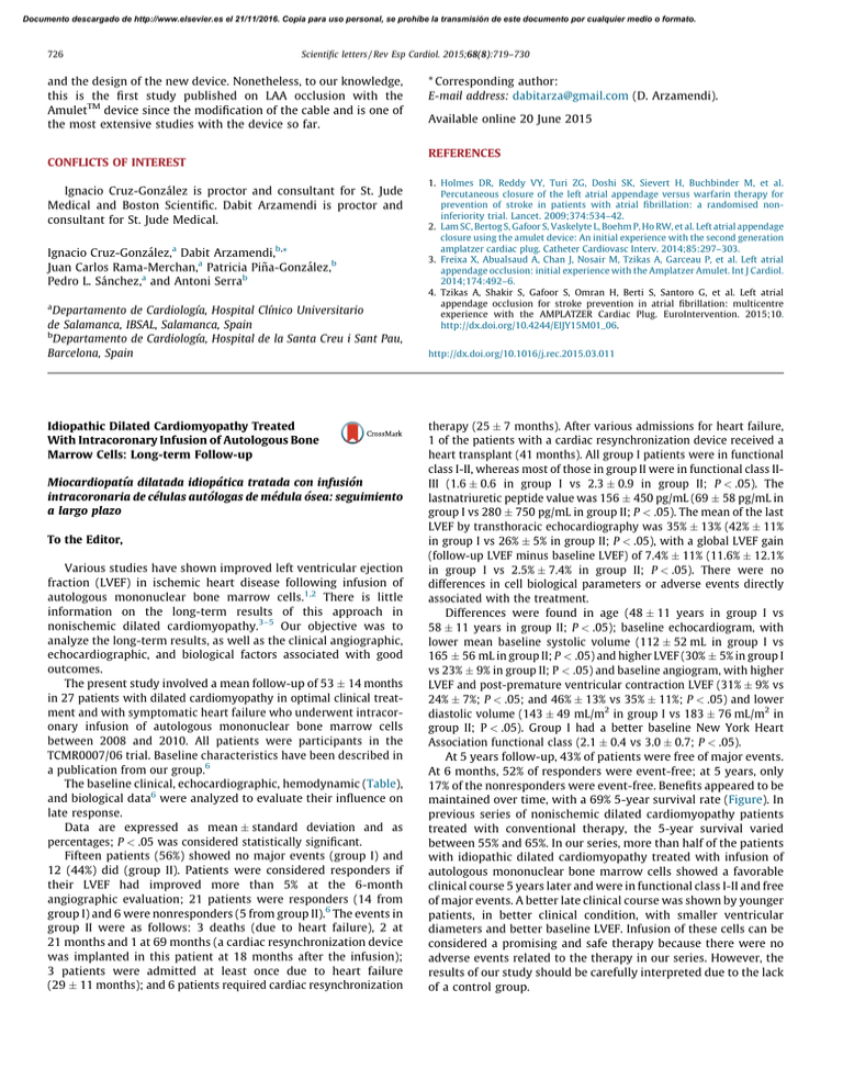
Documento descargado de http://www.elsevier.es el 21/11/2016. Copia para uso personal, se prohíbe la transmisión de este documento por cualquier medio o formato. 726 Scientific letters / Rev Esp Cardiol. 2015;68(8):719–730 and the design of the new device. Nonetheless, to our knowledge, this is the first study published on LAA occlusion with the AmuletTM device since the modification of the cable and is one of the most extensive studies with the device so far. CONFLICTS OF INTEREST Ignacio Cruz-González is proctor and consultant for St. Jude Medical and Boston Scientific. Dabit Arzamendi is proctor and consultant for St. Jude Medical. Ignacio Cruz-González,a Dabit Arzamendi,b,* Juan Carlos Rama-Merchan,a Patricia Piña-González,b Pedro L. Sánchez,a and Antoni Serrab a Departamento de Cardiologı´a, Hospital Clı´nico Universitario de Salamanca, IBSAL, Salamanca, Spain b Departamento de Cardiologı´a, Hospital de la Santa Creu i Sant Pau, Barcelona, Spain Idiopathic Dilated Cardiomyopathy Treated With Intracoronary Infusion of Autologous Bone Marrow Cells: Long-term Follow-up Miocardiopatı´a dilatada idiopática tratada con infusión intracoronaria de células autólogas de médula ósea: seguimiento a largo plazo To the Editor, Various studies have shown improved left ventricular ejection fraction (LVEF) in ischemic heart disease following infusion of autologous mononuclear bone marrow cells.1,2 There is little information on the long-term results of this approach in nonischemic dilated cardiomyopathy.3–5 Our objective was to analyze the long-term results, as well as the clinical angiographic, echocardiographic, and biological factors associated with good outcomes. The present study involved a mean follow-up of 53 14 months in 27 patients with dilated cardiomyopathy in optimal clinical treatment and with symptomatic heart failure who underwent intracoronary infusion of autologous mononuclear bone marrow cells between 2008 and 2010. All patients were participants in the TCMR0007/06 trial. Baseline characteristics have been described in a publication from our group.6 The baseline clinical, echocardiographic, hemodynamic (Table), and biological data6 were analyzed to evaluate their influence on late response. Data are expressed as mean standard deviation and as percentages; P < .05 was considered statistically significant. Fifteen patients (56%) showed no major events (group I) and 12 (44%) did (group II). Patients were considered responders if their LVEF had improved more than 5% at the 6-month angiographic evaluation; 21 patients were responders (14 from group I) and 6 were nonresponders (5 from group II).6 The events in group II were as follows: 3 deaths (due to heart failure), 2 at 21 months and 1 at 69 months (a cardiac resynchronization device was implanted in this patient at 18 months after the infusion); 3 patients were admitted at least once due to heart failure (29 11 months); and 6 patients required cardiac resynchronization * Corresponding author: E-mail address: dabitarza@gmail.com (D. Arzamendi). Available online 20 June 2015 REFERENCES 1. Holmes DR, Reddy VY, Turi ZG, Doshi SK, Sievert H, Buchbinder M, et al. Percutaneous closure of the left atrial appendage versus warfarin therapy for prevention of stroke in patients with atrial fibrillation: a randomised noninferiority trial. Lancet. 2009;374:534–42. 2. Lam SC, Bertog S, Gafoor S, Vaskelyte L, Boehm P, Ho RW, et al. Left atrial appendage closure using the amulet device: An initial experience with the second generation amplatzer cardiac plug. Catheter Cardiovasc Interv. 2014;85:297–303. 3. Freixa X, Abualsaud A, Chan J, Nosair M, Tzikas A, Garceau P, et al. Left atrial appendage occlusion: initial experience with the Amplatzer Amulet. Int J Cardiol. 2014;174:492–6. 4. Tzikas A, Shakir S, Gafoor S, Omran H, Berti S, Santoro G, et al. Left atrial appendage occlusion for stroke prevention in atrial fibrillation: multicentre experience with the AMPLATZER Cardiac Plug. EuroIntervention. 2015;10. http://dx.doi.org/10.4244/EIJY15M01_06. http://dx.doi.org/10.1016/j.rec.2015.03.011 therapy (25 7 months). After various admissions for heart failure, 1 of the patients with a cardiac resynchronization device received a heart transplant (41 months). All group I patients were in functional class I-II, whereas most of those in group II were in functional class IIIII (1.6 0.6 in group I vs 2.3 0.9 in group II; P < .05). The lastnatriuretic peptide value was 156 450 pg/mL (69 58 pg/mL in group I vs 280 750 pg/mL in group II; P < .05). The mean of the last LVEF by transthoracic echocardiography was 35% 13% (42% 11% in group I vs 26% 5% in group II; P < .05), with a global LVEF gain (follow-up LVEF minus baseline LVEF) of 7.4% 11% (11.6% 12.1% in group I vs 2.5% 7.4% in group II; P < .05). There were no differences in cell biological parameters or adverse events directly associated with the treatment. Differences were found in age (48 11 years in group I vs 58 11 years in group II; P < .05); baseline echocardiogram, with lower mean baseline systolic volume (112 52 mL in group I vs 165 56 mL in group II; P < .05) and higher LVEF (30% 5% in group I vs 23% 9% in group II; P < .05) and baseline angiogram, with higher LVEF and post-premature ventricular contraction LVEF (31% 9% vs 24% 7%; P < .05; and 46% 13% vs 35% 11%; P < .05) and lower diastolic volume (143 49 mL/m2 in group I vs 183 76 mL/m2 in group II; P < .05). Group I had a better baseline New York Heart Association functional class (2.1 0.4 vs 3.0 0.7; P < .05). At 5 years follow-up, 43% of patients were free of major events. At 6 months, 52% of responders were event-free; at 5 years, only 17% of the nonresponders were event-free. Benefits appeared to be maintained over time, with a 69% 5-year survival rate (Figure). In previous series of nonischemic dilated cardiomyopathy patients treated with conventional therapy, the 5-year survival varied between 55% and 65%. In our series, more than half of the patients with idiopathic dilated cardiomyopathy treated with infusion of autologous mononuclear bone marrow cells showed a favorable clinical course 5 years later and were in functional class I-II and free of major events. A better late clinical course was shown by younger patients, in better clinical condition, with smaller ventricular diameters and better baseline LVEF. Infusion of these cells can be considered a promising and safe therapy because there were no adverse events related to the therapy in our series. However, the results of our study should be carefully interpreted due to the lack of a control group. Documento descargado de http://www.elsevier.es el 21/11/2016. Copia para uso personal, se prohíbe la transmisión de este documento por cualquier medio o formato. Scientific letters / Rev Esp Cardiol. 2015;68(8):719–730 727 Table Baseline Clinical and Echocardiographic Parameters Group I (n = 15) Group II (n = 12) P Age, mean SD, y 49 12 58 11 <.05 Men, % 73 75 NYHA 2.2 0.4 2.7 0.7 NS <.05 BNP, pg/mL 341 415 481 424 NS Cholesterol, mg% 167 47 183 28 NS LDL-C, mg% 99 30 115 19 NS HDL-C, mg% 37 10 36 13 NS Triglycerides, mg% 145 107 168 108 NS Hypertension, % 53 58 NS Hyperlipidemia, % 60 75 NS Smoking, % 73 58 NS Diabetes mellitus, % 27 50 LVEF, % 30 5 23 9 NS <.05 LV end-diastolic volume, mL 164 78 214 64 NS LV end-systolic volume, mL 112 52 165 56 <.05 Degree of mitral regurgitation 0.53 0.7 0.50 0.5 LVEF in sinus rhythm, % 31 9 24 7 <.05 NS LVEF in sinus rhythm post-PVC, % 46 13 35 11 <.05 Sinus end-diastolic volume, mL/m2 143 49 183 76 <.05 Post-PVC end-diastolic volume, mL/m2 163 48 194 74 NS End-systolic sinus volume, mL/m2 99 41 142 64 NS Post-PVC end-systolic volume, mL/m2 90 40 127 60 NS Systolic pressure of the pulmonary artery, mmHg 34 18 37 13 NS BNP, brain natriuretic peptide; HDL-C, high-density lipoprotein cholesterol; LDL-C, low-density lipoprotein cholesterol; LV, left ventricle; LVEF, LV ejection fraction; NS, no significant; NYHA, New York Heart Association functional class; post-PVC, post-premature ventricular contraction. All echocardiographic parameters were determined using the Simpson method. Group I: patients without follow-up events. Group II: patients with follow-up events. Survival 100 100 75 75 50 43% 25 0 Probability, % Probability, % Event-free survival 69% 50 25 0 12 24 36 48 60 72 0 0 12 Months 24 36 48 60 72 Months Figure. Probability of event-free survival, 5-year survival, and changes in left ventricular ejection fraction at 5 years. Flor Baeza Garzón,a,* Miguel Romero,a Javier Suárez de Lezo,a Soledad Ojeda Pineda,a Concepción Herrera,b and José Suárez de Lezoa a Servicio de Cardiologı´a, Hospital Universitario Reina Sofı´a, Córdoba, Spain b Servicio de Hematologı´a, Hospital Universitario Reina Sofı´a, Córdoba, Spain * Corresponding author: E-mail address: flor-85@hotmail.es (F. Baeza Garzón). Available online 22 June 2015 REFERENCES 1. Strauer BE, Steinhoff G. 10 years of intracoronary and intramyocardial bone marrow stem cell therapy of the heart: from de methodological origin to clinical practice. J Am Coll Cardiol. 2011;58:1095–104. 2. Suárez de Lezo J, Herrera C, Romero MA, Pan M, Jiménez R, Carmona D, et al. Recuperación funcional tras infusión intracoronaria de células mononucleadas de médula ósea autóloga en pacientes con infarto crónico anterior y depresión severa de la función ventricular. Rev Esp Cardiol. 2010;63:1127–35. 3. Vrtovec B, Poglajen G, Sever M, Lezaic L, Domanovic D, Cernelc P, et al. Effects of intracoronary stem cell transplantation in patients with dilated cardiomyopathy. J Card Fail. 2011;17:272–81. 4. Vrtovec B, Poglajen G, Lezic L, Sever M, Domanovic D, Cernelc P, et al. Effects of intracoronary CD34+ stem cell transplantation in nonischemic dilated cardiomyopathy patients: 5-year follow-up. Circ Res. 2013;112:165–73. Documento descargado de http://www.elsevier.es el 21/11/2016. Copia para uso personal, se prohíbe la transmisión de este documento por cualquier medio o formato. Scientific letters / Rev Esp Cardiol. 2015;68(8):719–730 728 5. Seth S, Bhargava B, Narang R, Ray R, Mohanty S, Gulati G, et al.; AIIMS Stem Cell Study Group. The ABCD (Autologous Bone Marrow Cells in Dilated Cardiomyopathy) trial — A long-term follow-up study. J Am Coll Cardiol. 2010;55:1643–4. 6. Suárez de Lezo J, Herrera C, Romero M, Pan M, Suárez de Lezo Jr, Carmona MD, et al. Mejorı́a funcional en pacientes con miocardiopatı́a dilatada tras la infusión Long-term Results of Repeat Percutaneous Mitral Valvuloplasty: Is it Still a Viable Option? Resultados a largo plazo de la revalvuloplastia mitral percutánea: es todavı´a una opción real? ? To the Editor, In 1984, the percutaneous mitral valvuloplasty (PMV) technique of the Japanese surgeon Inoue was published, revolutionizing mitral stenosis treatment and, due to better results, supplanting the surgical technique.1 In the Euro Heart Survey of 2001, mitral stenosis was seen in 9.5% of 5001 patients.2 Of 112 patients who underwent a stenosis intervention, 34% received a percutaneous treatment.2 This intervention showed excellent results in subsequent studies with follow-up durations of up to 20 years.3 However, some valve restenosis patients later require valve replacement surgery; nonetheless, some patients could still be candidates for a repeat PMV. Because repeat PMV data are intracoronaria de células mononucleares autólogas de médula ósea. Rev Esp Cardiol. 2013;66:450–7. http://dx.doi.org/10.1016/j.rec.2015.03.012 scarce in the literature3 and nonexistent in Spain, our objective was to determine the characteristics and clinical course of patients who underwent a repeat PMV in an extensive PMV series. A total of 1138 consecutive PMV were retrospectively reviewed. These procedures were performed between 1988 and 2004 using the Inoue technique (with a single balloon containing 3 regions, initially in the form of an hourglass, the waist of the balloon is the least compliant part and opens the commissures). Clinical data were collected from the medical records and follow-up in the clinic or by telephone. Of the PMV, 35 repeat PMV were identified between 1989 and 2012. Of these, 5 were in men, and the mean patient age was 57.3 years (Table). Ten patients received a suboptimal PMV that required a second catheterization before 60 days. Overall, the median time to repeat PMV was 4.7 years (7.1 years if suboptimal PMV were excluded). The median followup was 10.8 years (Figure). One patient required urgent surgery after the repeat PMV due to procedure-related complications. During follow-up, 17 patients died (median survival, 8.1 years), 7 of cardiac causes (including 1 cardiac arrest and 2 related to Table General Characteristics of Patients in the Entire Cohort and Divided According to the Intervention Type (Elective or Due to a Previous Failed/Suboptimal Valvuloplasty). The Events Registered During Follow-up Are Detailed According to the Above Groups Total Elective Previous suboptimal valvuloplastya Patients 35 25 10 Age at 1st valvuloplasty, years 52.4 13.5 54.5 13.7 47.9 12.2 Age at repeat, years 57.3 13.8 61.3 12.7 47.9 12.2 Women 30 (85.7) 22 (88.0) 8 (80.0) Previous stroke 2 (5.7) 2 (8.0) 0 Chronic atrial fibrillation 17 (48.5) 14 (56.0) 3 (30.0) Previous surgical commissurotomy 4 (11.4) 4 (16.0) 0 Wilkins score 7.86 1.8 8.00 2.0 7.5 1.0 LVEF, % 64.5 7.1 64.5 7.9 63.4 3.9 Mitral gradient before repeat 12.0 (6.0-15.0) 11.5 (6.0-10.0) 12.0 (4.5-19.0) Mitral gradient after repeat 5.4 (3.9-7.5) 5.78 (3.9-8.0) 5.0 (2.7-7.2) Follow-up, years 10.8 [6.6-13.8] 10.2 [6.1-13.0] 16.9 [9.74-23.4] Urgent surgery (< 24 h) 1 (2.8) 0 1 (10.0) Mitral valve surgery 17 (48.5) 12 (48.0) 5 (50.0) Time to mitral surgery, yearsb 8.4 [2.0-13.1] 6.9 [1.52-12.1] 13.05 [4.31-23.4] Follow-up/preoperative de novo AF 4 (2.8)/17 (48.5) 3 (12.0)/14 (56.0) 1 (10.0)/3 (30.0) Stroke during follow-up 5 (14.2) 4 (16.0) 1 (10.0) Endocarditis 1 (2.8) 1 (4.0) 0 Readmission due to stable angina 1 (2.8) 0 1 (10.0) Acute coronary syndrome 2 (5.7) 2 (8.0) 0 Cardiogenic shock 2 (5.7) 2 (8.0) 0 NYHA > I 15 (42.8) 12 (48.0) 5 (50.0) MACE 26 (74.2) 20 (90.9) 6 (60.0) Death 17 (48.5) 14 (56.0) 3 (30.0) Events during follow-up AF, atrial fibrillation; LVEF, left ventricular ejection fraction; MACE, major adverse cardiac events (composite variable of death from any cause, mitral valve surgery, stroke, or mitral valve endocarditis); NYHA, New York Heart Association functional class. The data are expressed as No. (%), mean standard deviation, or median [interquartile range]. a Less than 2 months following the first procedure. b Considering the time from the first cardiac surgery or until the end of follow-up in those not requiring surgery, previously censored (n = 18; about 51% of patients).
